

Influence of bone lesion location on femoral bone strength assessed by MRI-based finite-element modeling
- PMID: 30851438
- PMCID: PMC6486650
- DOI: 10.1016/j.bone.2019.03.005
Influence of bone lesion location on femoral bone strength assessed by MRI-based finite-element modeling
Abstract
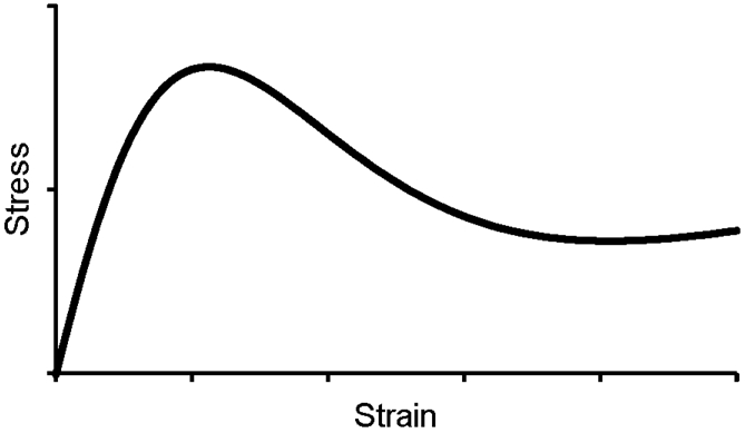
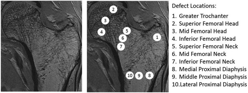
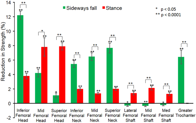
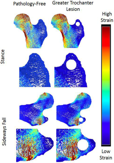
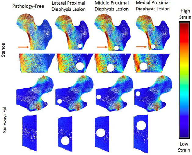
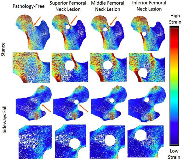
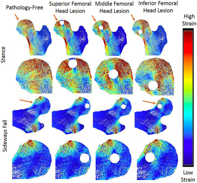
Currently, clinical determination of pathologic fracture risk in the hip is conducted using measures of defect size and shape in the stance loading condition. However, these measures often do not consider how changing lesion locations or how various loading conditions impact bone strength. The goal of this study was to determine the impact of defect location on bone strength parameters in both the sideways fall and stance-loading conditions. We recruited 20 female subjects aged 48-77 years for this study and performed MRI of the proximal femur. Using these images, we simulated 10-mm pathologic defects in greater trochanter, superior, middle, and inferior femoral head, superior, middle, and inferior femoral neck, and lateral, middle, and medial proximal diaphysis to determine the effect of defect location on change in bone strength by performing finite element analysis. We compared the effect of each osteolytic lesion on bone stiffness, strength, resilience, and toughness. For the sideways fall loading, defects in the inferior femoral head (12.21%) and in the greater trochanter (6.43%) resulted in the greatest overall reduction in bone strength. For the stance loading, defects in the mid femoral head (-7.91%) and superior femoral head (-7.82%) resulted in the greatest overall reduction in bone strength. Changes in stiffness, yield force, ultimate force, resilience, and toughness were not found to be significantly correlated between the sideways fall and stance-loading for the majority of defect locations, suggesting that calculations based on the stance-loading condition are not predictive of the change in bone strength experienced in the sideways fall condition. While stiffness was significantly related to yield force (R2 > 0.82), overall force (R2 > 0.59), and resilience (R2 > 0.55), in both, the stance-loading and sideways fall conditions for most defect locations, stiffness was not significantly related to toughness. Therefore, structure-dependent measure such as stiffness may not fully explain the post-yield measures, which depend on material failure properties. The data showed that MRI-based models have the sensitivity to determine the effect of pathologic lesions on bone strength.
Keywords: Lesion location; Proximal femur; Sideways fall; Stance; Stiffness; Strength.
Copyright © 2019 Elsevier Inc. All rights reserved.
Figures
Similar articles
-
In Vivo Assessment of Age- and Loading Configuration-Related Changes in Multiscale Mechanical Behavior of the Human Proximal Femur Using MRI-Based Finite Element Analysis.J Magn Reson Imaging. 2021 Mar;53(3):905-912. doi: 10.1002/jmri.27403. Epub 2020 Oct 19. J Magn Reson Imaging. 2021. PMID: 33075178
-
Study of stress variations in single-stance and sideways fall using image-based finite element analysis.Biomed Mater Eng. 2016 May 12;27(1):1-14. doi: 10.3233/BME-161563. Biomed Mater Eng. 2016. PMID: 27175463
-
Physical activity induced adaptation can increase proximal femur strength under loading from a fall onto the greater trochanter.Bone. 2021 Nov;152:116090. doi: 10.1016/j.bone.2021.116090. Epub 2021 Jun 25. Bone. 2021. PMID: 34175500 Free PMC article.
-
MRI-based assessment of proximal femur strength compared to mechanical testing.Bone. 2020 Apr;133:115227. doi: 10.1016/j.bone.2020.115227. Epub 2020 Jan 9. Bone. 2020. PMID: 31926345 Free PMC article.
-
Micro-Finite Element Analysis of the Proximal Femur on the Basis of High-Resolution Magnetic Resonance Images.Curr Osteoporos Rep. 2018 Dec;16(6):657-664. doi: 10.1007/s11914-018-0481-5. Curr Osteoporos Rep. 2018. PMID: 30232586 Free PMC article. Review.
Cited by
-
Fracture Risk Evaluation of Bone Metastases: A Burning Issue.Cancers (Basel). 2021 Nov 15;13(22):5711. doi: 10.3390/cancers13225711. Cancers (Basel). 2021. PMID: 34830865 Free PMC article. Review.
-
Predicting Fractures Using Vertebral 18F-NaF Uptake in Prostate Cancer Patients.J Bone Metab. 2023 Nov;30(4):329-337. doi: 10.11005/jbm.2023.30.4.329. Epub 2023 Nov 30. J Bone Metab. 2023. PMID: 38073266 Free PMC article.
-
The effect of variations in CT scan protocol on femoral finite element failure load assessment using phantomless calibration.PLoS One. 2022 Mar 18;17(3):e0265524. doi: 10.1371/journal.pone.0265524. eCollection 2022. PLoS One. 2022. PMID: 35303026 Free PMC article.
-
Two Cannulated Screws Provide Sufficient Biomechanical Strength for Prophylactic Fixation in Adult Patients With an Aggressive Benign Femoral Neck Lesion.Front Bioeng Biotechnol. 2022 Jul 7;10:891338. doi: 10.3389/fbioe.2022.891338. eCollection 2022. Front Bioeng Biotechnol. 2022. PMID: 35875489 Free PMC article.
-
Patient-Specific Bone Multiscale Modelling, Fracture Simulation and Risk Analysis-A Survey.Materials (Basel). 2019 Dec 24;13(1):106. doi: 10.3390/ma13010106. Materials (Basel). 2019. PMID: 31878356 Free PMC article. Review.
References
-
- Hipp JA, Springfield DS, and Hayes WC, Predicting pathologic fracture risk in the management of metastatic bone defects. Clin Orthop Relat Res, 1995(312): p. 120–35. - PubMed
-
- Hipp JA, et al., Trabecular bone morphology from micro-magnetic resonance imaging. J Bone Miner Res, 1996. 11(2): p. 286–97. - PubMed
-
- Toomey A and Friedman L, Mortality in cancer patients after a fall-related injury: The impact of cancer spread and type. Injury, 2014. 45(11): p. 1710–6. - PubMed
-
- Crowninshield RD, et al., A biomechanical investigation of the human hip. J Biomech, 197811(1-2): p. 75–85. - PubMed
-
- Lotz JC, Cheal EJ, and Hayes WC, Stress distributions within the proximal femur during gait and falls: implications for osteoporotic fracture. Osteoporos Int, 1995. 5(4): p. 252–61. - PubMed
Publication types
MeSH terms
Grants and funding
LinkOut - more resources
Full Text Sources
Medical
