. 2019 Apr 18;74(2):310-319.e7.
doi: 10.1016/j.molcel.2019.01.043. Epub 2019 Mar 6.

The Ribosome Cooperates with a Chaperone to Guide Multi-domain Protein Folding

Affiliations

The Ribosome Cooperates with a Chaperone to Guide Multi-domain Protein Folding

Kaixian Liu et al. Mol Cell. .

Abstract

Multi-domain proteins, containing several structural units within a single polypeptide, constitute a large fraction of all proteomes. Co-translational folding is assumed to simplify the conformational search problem for large proteins, but the events leading to correctly folded, functional structures remain poorly characterized. Similarly, how the ribosome and molecular chaperones promote efficient folding remains obscure. Using optical tweezers, we have dissected early folding events of nascent elongation factor G, a multi-domain protein that requires chaperones for folding. The ribosome and the chaperone trigger factor reduce inter-domain misfolding, permitting folding of the N-terminal G-domain. Successful completion of this step is a crucial prerequisite for folding of the next domain. Unexpectedly, co-translational folding does not proceed unidirectionally; emerging unfolded polypeptide can denature an already-folded domain. Trigger factor, but not the ribosome, protects against denaturation. The chaperone thus serves a previously unappreciated function, helping multi-domain proteins overcome inherent challenges during co-translational folding.

Keywords: elongation factor G; molecular chaperones; multi-domain protein; nascent polypeptide; optical tweezers; protein folding; protein synthesis; ribosome; single-molecule; translation.


Conflict of interest statement

DECLARATION OF INTERESTS.

The authors declare no competing interests.

Figures

Fig. 1
Fig. 1. The ribosome modulates G-domain folding.
A. Experimental scheme. The bar diagram on top illustrates the EF-G domain topology. For optical tweezers experiments, stalled ribosome-nascent chain complexes (RNCs), generated in vitro using stop codon-less mRNAs, are tethered between two polystyrene beads. B. Example force-extension curves (FECs) for 328RNC. The native G-domain unfolds in the initial FEC (red trace). In subsequent pulls (black), separated by 10 second refolding pauses, unfolding transitions from the fully structured state are apparent in some of the traces. In addition, partially structured and molten globule-like states are observed. C. Apparent folding rates of the G-domain for the isolated Galone polypeptide (top, grey bar) and RNCs with varying lengths (brown bars). The cartoons on the right indicate the stalling positions for the respective constructs. The ribosome decelerates folding at short nascent chain lengths (328RNC vs. Galone). After reaching a maximum, folding rates drop in longer nascent chains due to non-productive interactions within the nascent polypeptide. This interference is even more pronounced in the isolated G-II protein comprised of the G-domain and domain II (bottom, grey bar), indicating that the ribosome reduces non-productive inter-domain interactions.
Fig. 2
Fig. 2. The ribosome and trigger factor reduce inter-domain misfolding.
A. Example of G-domain refolding against a constant force of 3.5 pN for G-II. Raw data (1 kHz) is shown as grey dots, time-averaged data (10 Hz) as a line (orange before G-domain folding, black thereafter). Black dashed lines indicate states before and after G-domain folding (GU-IIU, GN-IIU). G-II also populates a misfolded state with a shorter extension (magenta dashed line). Histogram (right): extensions populated before (orange) and after (black) G-domain folding. B. Close-up view of the refolding transitions for four G-II example traces. In all cases, the misfolded state partially unfolds before productive folding occurs (black and magenta dashed lines as in A). C. Constant force refolding of 452RNC. The population of the compact misfolded species is reduced, as apparent from the extension-vs.-time trace and the extension histogram. D. Refolding in the presence of TF. The chaperone shifts the population away from the misfolded state. E. Aggregated extension histograms for the equilibrium portion of constant force refolding traces, aligned to the GN-IIU state. The misfolded state is highly populated in G-II (orange), but less so in 452RNC without (brown) and with (green) TF. F. Apparent G-domain refolding rate for G-II, 452RNC (as in Fig. 1C) and 452RNC + TF. Reduced misfolding in the presence of the ribosome and the chaperone results in faster folding.
Fig. 3
Fig. 3. Contacts with the G-domain stabilize domain II.
A. Example FECs for G-II. Clear signatures of both domains (colored arrowheads) are apparent in the initial trace (black). The G-domain refolds in some of the attempts (black arrowheads), but refolding of domain II is not observed subsequent traces (grey). B. Transient population of an unfolding intermediate (arrowhead) during II unfolding. Inset: magnified view of the unfolding transition. The short lifetime of the intermediate indicates that it is unstable. C. Schematic illustrating the likely pathway for two-step unfolding of domain II in G-II. The interface between the G-domain and domain II breaks first, followed by unfolding of the remaining domain II structure. The table lists the contour length changes expected based on the EF-G structure (“calc.”) and the observed values (“obs”) for the transitions. D. Scatter plot of domain II unfolding transitions in G-II. Each dot represents an unfolding transition. Transitions from two-step unfolding via the intermediate are connected by dotted lines. Dashed and solid lines represent WLC models calculated for two-step (ΔLN-I, ΔLI-U) and one-step (ΔLN-U) unfolding (see panel C). The shaded areas indicate the respective standard deviations of the observed contour length changes.
Fig. 4
Fig. 4. Domain II folding requires the folded G-domain.
A. Example FECs showing slow folding of domain II. Domain II is selectively unfolded while leaving the G-domain structured (yellow trace, “initial”). Refolding is apparent in trace “9” (yellow) after several futile attempts (black traces, “1-8”). Subsequent forced unfolding confirms domain II refolding (yellow trace, “probe”). Traces are plotted with an offset along the x axis for clarity, the “initial” trace is re-plotted in white as a reference. B. Scatter plot for unfolding of renatured domain II, confirming folding to the native structure in the presence of the folded G-domain. Dashed and solid yellow lines represent WLC models for two-step and one-step unfolding, shaded regions indicate the standard deviation of the experimental data. C. Domain II does not stably fold by itself because it requires stabilizing contacts at the interface with the G-domain. Consequently, the G-domain has to fold first, imposing a hierarchical folding order.
Fig. 5
Fig. 5. Unfolded domain II destabilizes the native G-domain.
A. Example of G-domain denaturation. After selectively unfolding domain II (yellow, “initial”), unfolding of the G-domain at low force during the second cycle (magenta, “2”) indicates denaturation. The initial trace is re-plotted in white as a reference. B. Distribution of unfolding forces. The native G-domain (red) unfolds in a force range that is well separated from that after denaturation (magenta). C. Scatter plot showing individual unfolding events after G-domain denaturation (magenta). The distribution resembles that of molten globule-like and partially structured states observed after mechanical unfolding of 328RNC (grey dots, data from fig. S2E), indicating that denaturation ultimately results in complete unfolding. D. Cumulative probability of domain II refolding (yellow) and G-domain denaturation (magenta) during repeated force ramp cycles. Both events occur with similar probability and thus on similar timescales. E. In addition to folding productively, domain II can cause G-domain denaturation, which then results in complete unfolding of the polypeptide. The two processes compete with each other.
Fig. 6
Fig. 6. TF protects against denaturation.
A. Cumulative probability distributions of G-domain denaturation. The ribosome does not protect against denaturation (G-II vs. 452RNC), which already occurs after partial synthesis of domain II (386RNC). TF blocks denaturation (452RNC + TF). B. Before domain II has been fully synthesized (386RNC), it cannot fold, but already destabilizes the neighboring folded G-domain. After domain II has emerged from the ribosome (452RNC), competing pathways result in either G-domain denaturation or folding of domain II. TF blocks denaturation, favoring folding.
Fig. 7
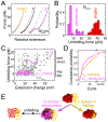
Fig. 7. Folding events during EF-G synthesis.
After the G-domain has emerged from the ribosome (top), its folding rate is reduced by interactions with the ribosome. Presumably similar interactions, now with domain II, reduce inter-domain misfolding in longer nascent chains (bottom), effectively increasing the G-domain folding rate. How the ribosome affects a particular domain thus depends on nascent chain length. The reduction in inter-domain misfolding is reinforced by TF. G-domain folding has to precede folding of domain II. Domain II folding competes with denaturation of the already folded G-domain. The latter process is prevented by TF, but not the ribosome. Hence, TF has a dual function in promoting early folding events in nascent EF-G.

Similar articles

Cited by

References

    1. AEvarsson A, Brazhnikov E, Garber M, Zheltonosova J, Chirgadze Y, al-Karadaghi S, Svensson LA, and Liljas A (1994). Three-dimensional structure of the ribosomal translocase: elongation factor G from Thermus thermophilus. The EMBO journal 13, 3669–3677. - PMC - PubMed
    1. Agashe VR, Guha S, Chang HC, Genevaux P, Hayer-Hartl M, Stemp M, Georgopoulos C, Hartl FU, and Barral JM (2004). Function of trigger factor and DnaK in multidomain protein folding: increase in yield at the expense of folding speed. Cell 117, 199–209. - PubMed
    1. Anfinsen CB (1973). Principles that govern the folding of protein chains. Science 181, 223–230. - PubMed
    1. Batey S, Nickson AA, and Clarke J (2008). Studying the folding of multidomain proteins. HFSP J 2, 365–377. - PMC - PubMed
    1. Beckett D, Kovaleva E, and Schatz PJ (1999). A minimal peptide substrate in biotin holoenzyme synthetase-catalyzed biotinylation. Protein Sci 8, 921–929. - PMC - PubMed

Publication types