. 2019 May:531:48-56.
doi: 10.1016/j.virol.2019.02.017. Epub 2019 Feb 26.

N-Linked glycosylation of the membrane protein ectodomain regulates infectious bronchitis virus-induced ER stress response, apoptosis and pathogenesis

Affiliations

N-Linked glycosylation of the membrane protein ectodomain regulates infectious bronchitis virus-induced ER stress response, apoptosis and pathogenesis

Jia Qi Liang et al. Virology. 2019 May.

Abstract

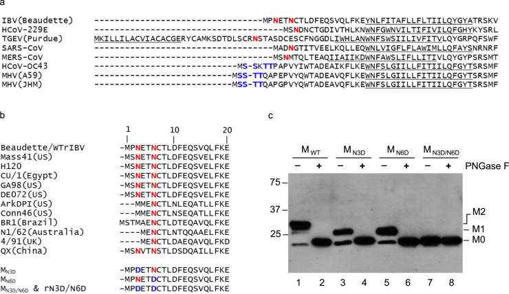
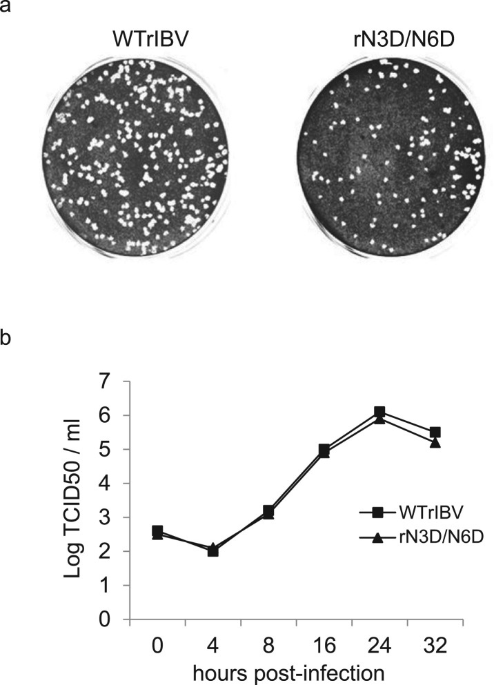
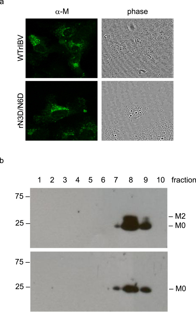
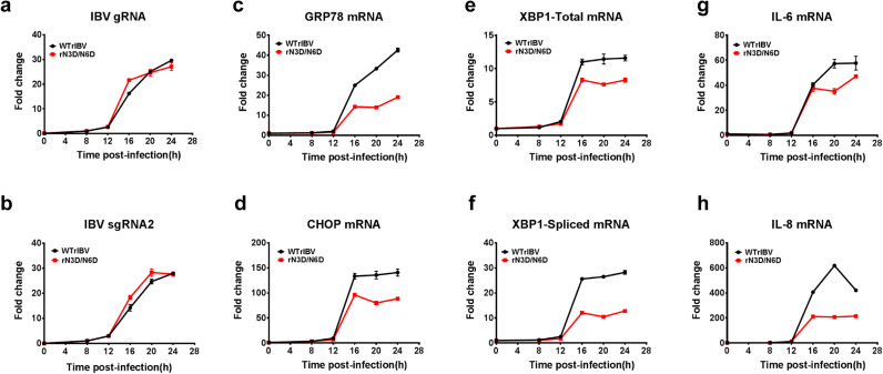
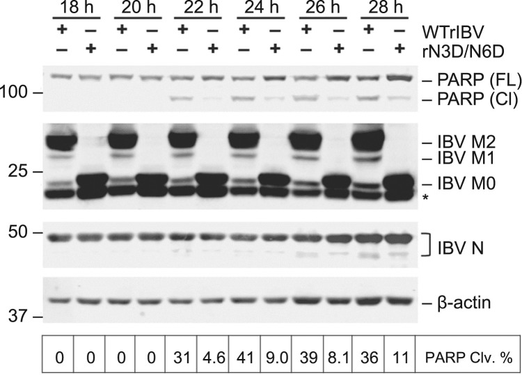
Coronavirus membrane (M) protein is the most abundant structural protein playing a critical role in virion assembly. Previous studies show that the N-terminal ectodomain of M protein is modified by glycosylation, but its precise functions are yet to be thoroughly investigated. In this study, we confirm that N-linked glycosylation occurs at two predicted sites in the M protein ectodomain of infectious bronchitis coronavirus (IBV). Dual mutations at the two sites (N3D/N6D) did not affect particle assembly, virus-like particle formation and viral replication in culture cells. However, activation of the ER stress response was significantly reduced in cells infected with rN3D/N6D, correlated with a lower level of apoptosis and reduced production of pro-inflammatory cytokines. Taken together, this study demonstrates that although not essential for replication, glycosylation in the IBV M protein ectodomain plays important roles in activating ER stress, apoptosis and proinflammatory response, and may contribute to the pathogenesis of IBV.

Keywords: Apoptosis; Coronavirus; ER stress; Glycosylation; Particle assembly; Pro-inflammatory response; Viral pathogenesis.


Figures

Fig. 1
Fig. 1
Alignment of the N-terminal sequences of coronavirus M proteins and the effects of glycosylation of IBV M protein on the protein expression. a. Alignment of the N-terminal sequences of coronavirus M proteins. The proposed transmembrane domains are underlined. Putative N-linked and O-linked glycosylation sites are indicated in red and blue, respectively. b. Alignment of the N-terminal sequences of M proteins in different IBV strains. The proposed transmembrane domains are underlined. Putative N-linked glycosylation sites are indicated in red. Generation of three mutants, MN3D, MN6D, and MN3D/N6D. Asn to Asp substitutions were introduced into the M protein to change the either one or both of the two predicted N-linked glycosylation sites at the amino acid positions 3 and 6, respectively. c. Expression and glycosylation of wild type and mutant M proteins. Total cell lysates prepared from HeLa cells expressing IBV MWT, MN3D, MN6D, and MN3D/N6D were either treated with or without PNGase F. Polypeptides were separated by SDS-PAGE and analyzed by Western blot with anti-IBV M antiserum. Numbers on the left indicate molecular masses in kilo Daltons. The unglycosylated (M0), glycosylated at one position (M1) and glycosylated at two positions (M2) forms of the M protein are indicated.
Fig. 2
Fig. 2
Plaque morphologies and growth kinetics of WTrIBV and the rN3D/N6D mutant. a. Plaque sizes and morphologies of WTrIBV and rN3D/N6D. Monolayers of Vero cells on a 6-well plate were infected with 100 µl of 1000-fold diluted virus stocks and cultured in the presence of 0.5% carboxymethyl cellulose at 37 °C for 3 days. Cells were then fixed and stained with 0.1% toluidine. b. The growth curves of WTrIBV and rN3D/N6D. Vero cells were infected with the viruses and harvested at 0, 4, 8, 16, 24 and 32 h post-inoculation, respectively. Viral stocks were prepared by freezing/thawing the cells three times, and Vero cells on 96-well plates were infected with 10-fold serial dilution of the viral stock. TCID50 was determined by using the Reed and Muench method.
Fig. 3
Fig. 3
The subcellular localization of M protein and the sedimentation properties of WTrIBV and the rN3D/N6D mutant. a. Intracellular distribution of M protein in cells infected with WTrIBV or rN3D/N6D. Vero cells were infected with WTrIBV or rN3D/N6D before being fixed, permeabilized and incubated with anti-IBV M antiserum. The bound primary antibodies were detected using FITC-labelled anti-rabbit secondary antibodies. b. Sedimental analysis of WTrIBV and the rN3D/N6D mutant. Vero cells were infected with WTrIBV or rN3D/N6D. Viral particles in the culture media were pelleted through 30% sucrose cushion and then sedimented into a discontinuous sucrose gradient consisting of 20%, 30%, 40%, 50% and 60% sucrose 2 ml each in TNE buffer. Ten fractions, 1 ml each, were collected from the top to the bottom for analysis by Western blot with anti-IBV M antiserum.
Fig. 4
Fig. 4
The effect of N3D/N6D mutation of IBV M protein on VLP assembly. COS-7 cells were infected with the recombinant vaccinia/T7 virus (vTF7-3) and transfected with a plasmid encoding wild type IBV E gene, together with a plasmid encoding MWT or MN3D/N6D, respectively. Intracellular cell lysates and pelleted extracellular VLPs were analyzed by Western blot with antisera against IBV M and E proteins.
Fig. 5
Fig. 5
Induction ER stress response and pro-inflammatory cytokines in cells infected with WTrIBV or the rN3D/N6D mutant. Vero cells were infected with WTrIBV or rN3D/N6D at MOI~2 and cellular RNA was harvested in a time course experiment. The amounts of IBV genomic RNA (gRNA), IBV subgenomic mRNA2 (sgRNA2), ER stress-related genes and two proinflammatory cytokines were determined by RT-qPCR, using GAPDH as an internal control. The experiment was repeated three times with similar results, and the result of one representative experiment is shown.
Fig. 6
Fig. 6
Induction of apoptosis in cells infected with WTrIBV or the rN3D/N6D mutant. Vero cells were infected with WTrIBV or rN3D/N6D at MOI~2. Protein lysates were harvested at 18 hpi onwards in 2 h intervals and were subjected to Western blot analysis using the indicated antisera or antibodies. Beta-actin was included as the loading control. Sizes of protein ladders in kilo Dalton were indicated on the left. Degree of cell apoptosis was calculated as the band intensity of cleaved PARP protein divided by the band intensity of the total protein. The experiment was repeated three times with similar results, and the result of one representative experiment is shown.
Fig. 7
Fig. 7
ELD50 of WTrIBV and the rN3D/N6D mutant. The virus stocks of WTrIBV and rN3D/N6D of the same titers were 10-fold serially diluted and 0.2 ml diluted virus was injected into the allantoic cavity of 10-day old embryonated SPF eggs and incubated at 37 °C for 5 days. The numbers of live or dead embryos were counted and the 50% embryo lethal dose (ELD50) was calculated using the Reed and Muench method. The bar chart shows the results from two independent experiments with standard deviations.

Similar articles

Cited by

References

    1. Bárcena M., Oostergetel G.T., Bartelink W., Faas F.G.A., Verkleij A., Rottier P.J.M., Koster A.J., Bosch B.J. Cryo-electron tomography of mouse hepatitis virus: insights into the structure of the coronavirion. Proc. Natl. Acad. Sci. USA. 2009;106:582–587. - PMC - PubMed
    1. Binns M.M., Boursnell M.E., Tomley F.M., Brown T.D. Nucleotide sequence encoding the membrane protein of the IBV strain 6/82. Nucleic Acids Res. 1986;14:5558. - PMC - PubMed
    1. Cavanagh D. Coronavirus avian infectious bronchitis virus. Vet. Res. 2007;38:281–297. - PubMed
    1. Cavanagh D., Davis P.J., Pappin D.J. Coronavirus IBV glycopolypeptides: locational studies using proteases and saponin, a membrane permeabilizer. Virus Res. 1986;4:145–156. - PMC - PubMed
    1. Collins M.S., Alexander D.J., Harkness J.W. Heterogeneity of infectious bronchitis virus grown in eggs. Arch. Virol. 1976;50:55–72. - PMC - PubMed

Publication types

MeSH terms

Substances