

Case Reports
doi: 10.1136/bcr-2018-227408.
Severe tricuspid valve destruction secondary to previous non-appropriately treated endocarditis
Affiliations
- PMID: 30852499
- PMCID: PMC6424179
- DOI: 10.1136/bcr-2018-227408
Item in Clipboard
Case Reports
Severe tricuspid valve destruction secondary to previous non-appropriately treated endocarditis
BMJ Case Rep.
.
Abstract
Tricuspid valve(TV) destruction with a remote history of endocarditis without known risk factors (ie, HIV, intravenous drug use, neoplasm, trauma) is rare. We describe the case of a TVs destruction in a 12-year-old non-HIV boy, with a 4-year history of endocarditis without known risk factors nor evidence regarding previous appropriately management.
Keywords: heart failure; valvar diseases.
© BMJ Publishing Group Limited 2019. No commercial re-use. See rights and permissions. Published by BMJ.
Conflict of interest statement
Competing interests: None declared.
Figures

Transthoracic echocardiogram. Four chamber view shows tricuspid valve (TV) endocarditis with large vegetation (arrow) on anterior and posterior pathological TVs. LA, left atrium; LV, left ventricle; RA, right atrium; RV, right ventricle.
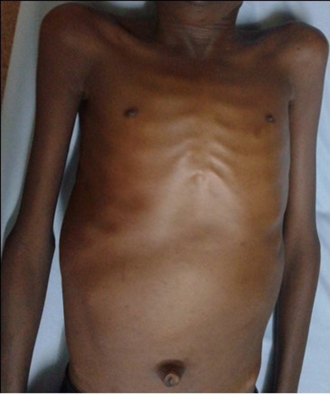
Clinical presentation of patient with visible hepatomegaly at inspection.

Chest radiograph shows severe cardiomegaly with right ventricular and right atrial dilation. CTI, cardiothoracic index.

ECG shows atrial fibrillation with ventricular aberrations, right axial deviation, right ventricular hypertrophy, complete right bundle branch block.

Transthoracic echocardiogram. Four-chamber view shows tricuspid valve (TV) remnants (white arrow) and marked right ventricular and right atrial dilation with severe tricuspid regurgitation secondary to TV destruction and annular dilatation (yellow arrow). LA, left atrium; LV, left ventricle; RA, right atrium; RV, right ventricle.
References
Publication types
MeSH terms
LinkOut - more resources
Full Text Sources
Medical