. 2019 Jul;256(4):1051-1063.
doi: 10.1007/s00709-019-01363-5. Epub 2019 Mar 9.

Distribution of plastids and mitochondria during male gametophyte formation in Tinantia erecta (Jacq.) Fenzl

Affiliations

Distribution of plastids and mitochondria during male gametophyte formation in Tinantia erecta (Jacq.) Fenzl

Rafał Marciniec et al. Protoplasma. 2019 Jul.

Abstract

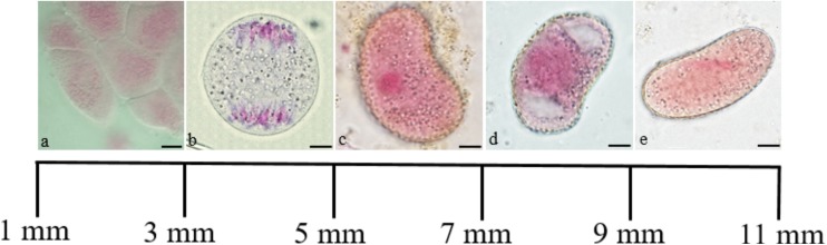
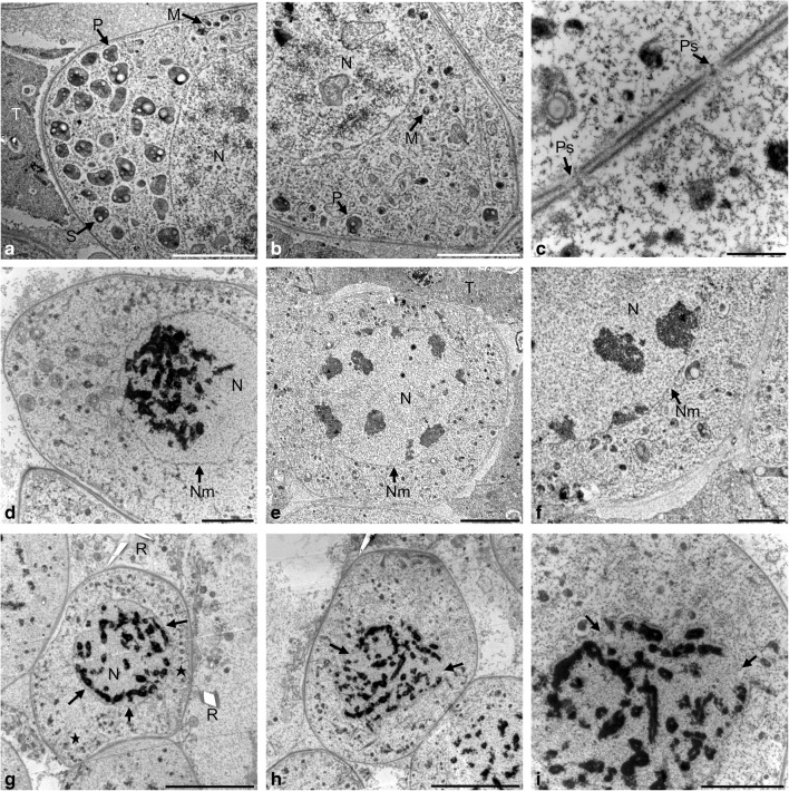
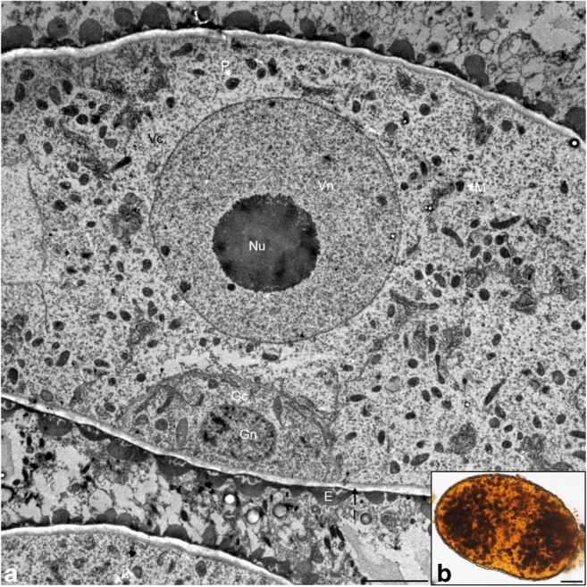
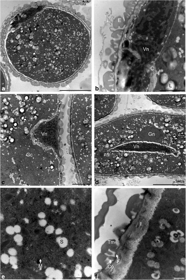
During meiosis in microsporogenesis, autonomous cellular organelles, i.e., plastids and mitochondria, move and separate into daughter cells according to a specific pattern. This process called chondriokinesis is characteristic for a given plant species. The key criterion for classification of the chondriokinesis types was the arrangement of cell organelles during two meiosis phases: metaphase I and telophase I. The autonomous organelles participate in cytoplasmic inheritance; therefore, their precise distribution to daughter cells determines formation of identical viable microspores. In this study, the course of chondriokinesis during the development of the male gametophyte in Tinantia erecta was analyzed. The study was conducted using optical and transmission electron microscopes. During microsporogenesis in T. erecta, autonomous cell organelles moved in a manner defined as a neutral-equatorial type of chondriokinesis. Therefore, metaphase I plastids and mitochondria were evenly dispersed around the metaphase plate and formed an equatorial plate between the daughter nuclei in early telophase I. Changes in the ultrastructure of plastids and mitochondria during pollen microsporogenesis were also observed.

Keywords: Chondriokinesis; Microgametogenesis; Microsporogenesis; Mitochondria; Plastids; Tinantia erecta.


Conflict of interest statement

The authors declare that they have no conflict of interest.

Figures

Fig. 1
Fig. 1
Relationship between the length of the flower bud and the stage of development of T. erecta male gametophyte observed in light microscopy (LM) on crushed preparations stained with acetocarmine. a Sporogenous tissue. b Anaphase I. c Free microspore. d Vacuolated microspore. e Binucleate pollen grain. Scale bars: 10 μm
Fig. 2
Fig. 2
Meiotic prophase I observed in TEM. a Grouping of plastids on the cell pole. b Distribution of organelles at the opposite pole of the microsporocyte. c Plasmodesmata between microsporocytes. d Concentration of chromatin in the bouquet stage in zygotene. e Pachytene. f Clusters of heterochromatin adjacent to the nuclear envelope. g Diplotene. h Diakinesis. i Disintegration of the nuclear membrane at the diakinese stage. Scale bars: 5 μm in a, b, e, g, h; 2 μm in d, f, i; 500 nm in c (M mitochondrion, N nucleus, Nm nuclear membrane, P plastid, Ps plasdodesmata, R raphide, S starch grain, T tapetum, Tn tapetal nucleus)
Fig. 3
Fig. 3
Metaphase I and anaphase I in meiotic cells observed in TEM. a Metaphase I. b Cell organelles surrounded by the endoplasmic reticulum network. c Early anaphase I. d Anaphase I. e Cell organelles arranged parallel to the wall of the microsporocyte. f Organelles grouped in a triangle. Scale bars: 5 μm in a, c, d; 2 μm in b, e, f (Ch chromosomes, M mitochondrion, Mf multimembranous formations, P plastid, R raphide, ER endoplasmic reticulum, T tapetum, Tn tapetal nucleus)
Fig. 4
Fig. 4
Telophase I and prophase II in meiotic cells. a Organelles grouped in triangles during telophase I, semi-thin section stained with toluidine blue. b Telophase I, TEM. c Vesicles forming the primary cell wall, TEM. d Prophase II, TEM. e Formed primary cell wall, TEM. f Clusters of starch in individual dyad cells, crushed preparation stained in IKI reaction. Scale bars: 10 μm in a, f; 5 μm in b, d; 1 μm in c, e (C callose, Ch chromosomes, Cw cell wall, M mitochondrion, N nucleus, Nm nuclear membrane, Nu nucleolus, P plastid, PCW primary cell wall, R raphides, ER endoplasmic reticulum, S starch grain, T tapetum)
Fig. 5
Fig. 5
Metaphase II and anaphase II in microsporocytes. a Parietal arrangement of cell organelles during metaphase II, TEM. b Organelle aggregate, TEM. c Early anaphase II, semi-thin preparation stained with toluidine blue. d Anaphase II, TEM. Scale bars: 5 μm in a; 2 μm in b, d; 10 μm in c (C callose, Ch chromosomes, Cw cell wall, M mitochondrion, P plastid, R raphide, ER endoplasmic reticulum, T tapetum, Tn tapetal nucleus)
Fig. 6
Fig. 6
Telophase II and tetrad stage in microspores. a Primary wall at the stage of telophase II, semi-thin preparation stained with toluidine blue. b, c Telophase II, TEM. d Tetrahedral arrangement of the microspores in the tetrad, TEM. e Alternating arrangement of the microspores in the tetrad, semi-thin preparation stained with toluidine blue. f Single microspore in the tetrad, TEM. Scale bars: 10 μm in a, e; 5 μm in b, d; 2 μm in c; 1 μm in f (C callose, Cw cell wall, M mitochondrion, N nucleus, Nm nuclear membrane, Nu nucleolus, P plastid, PCW primary cell wall, R raphide, T tapetum)
Fig. 7
Fig. 7
Microgametogenesis stage. a Microspore after the first mitotic division, TEM. b Starch grains in the microspore, crushed preparation stained in IKI reaction. Scale bars: 1 μm in a; 10 μm in b (E exine, Gc generative cell, Gn generative nucleus, I intine, M mitochondrion, Nu nucleolus, P plastid, Vc vegetative cell, Vn vegetative nucleus)
Fig. 8
Fig. 8
Pollen grain observed in TEM. a Parietal position of the generative nucleus. b Nucleus of the generative cell. c Generative cell detaching from the sporoderm. d Central arrangement of the vegetative and generative nucleus. e Ultrastructure of the cytoplasm of the pollen grain. f Ultrastructure of the pollen grain sporoderm. Scale bars: 5 μm in a, d; 1 μm in b; 2 μm in c; 250 nm in e, f (C callose, Co columella, E exine, F foot layer, Gc generative cell, Gn generative nucleus, I intine, L lipid body, M mitochondrion, P plastid, Vc vegetative cell, Vn vegetative nucleus, Tm tectum)

Similar articles

Cited by

References

    1. Baker HG, Baker I. Starch in angiosperm pollen grains and its evolutionary significance. Am J Bot. 1979;66:591–600. doi: 10.1002/j.1537-2197.1979.tb06262.x. - DOI
    1. Bąkowski Z. Versucheiner Klassifizierung der Chondriokinese bei Kormophyten. Acta Soc Bot Pol. 1938;15:323–369. doi: 10.5586/asbp.1938.020. - DOI
    1. Bass HW, Marshall WF, Sedat JW, Agard DA, Cande WZ. Telomeres cluster de novo before the initiation of synapsis: a three dimensional spatial analysis of telomere positions before and during meiotic prophase. J Cell Biol. 1997;137:5–18. doi: 10.1083/jcb.137.1.5. - DOI - PMC - PubMed
    1. Batygina TB. Cabombaceae, Nymphaeaceae. In: Yakovlev MS, editor. Comparative embryology of flowering plants: Winteraceae-Juglandaceae. Leningrad: Nauka; 1981. pp. 101–110.
    1. Bednara J, Rodkiewicz B (1988) Cytoplasmic organelles in microsporocytes of Larix and sporocytes of Polystichum. Ann Sci Univ Reims 23:51–53

LinkOut - more resources