. 2019 Jun:100:52-61.
doi: 10.1016/j.jaut.2019.02.006. Epub 2019 Mar 8.

AS101 ameliorates experimental autoimmune uveitis by regulating Th1 and Th17 responses and inducing Treg cells

Affiliations

AS101 ameliorates experimental autoimmune uveitis by regulating Th1 and Th17 responses and inducing Treg cells

So Jin Bing et al. J Autoimmun. 2019 Jun.

Abstract

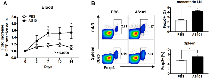
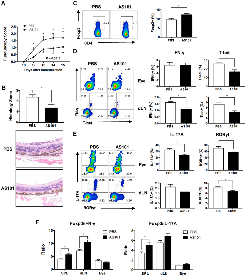
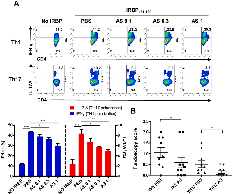
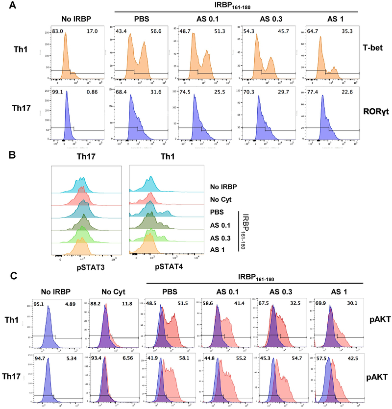
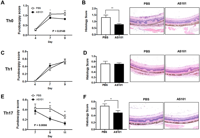
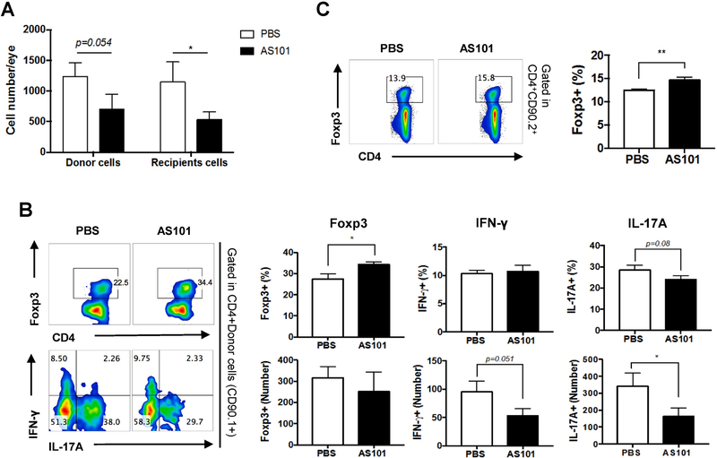
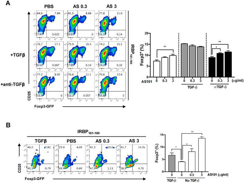
AS101 is an organotellurium compound with multifaceted immunoregulatory properties that is remarkable for its lack of toxicity. We tested the therapeutic effect of AS101 in experimental autoimmune uveitis (EAU), a model for human autoimmune uveitis. Unexpectedly, treatment with AS101 elicited Treg generation in vivo in otherwise unmanipulated mice. Mice immunized for EAU with the retinal antigen IRBP and treated with AS101 developed attenuated disease, as did AS101-treated recipients of retina-specific T cells activated in vitro. In both settings, eye-infiltrating effector T cells were decreased, whereas regulatory T (Treg) cells in the spleen were increased. Mechanistic studies in vitro revealed that AS101 restricted polarization of retina-specific T cells towards Th1 or Th17 lineage by repressing activation of their respective lineage-specific transcription factors and downstream signals. Retina-specific T cells polarized in vitro towards Th1 or Th17 in the presence of AS101 had impaired ability to induce EAU in naïve recipients. Finally, AS101 promoted differentiation of retina-specific T cells to Tregs in vitro independently of TGF-β. We conclude that AS101 modulates autoimmune T cells by inhibiting acquisition and expression of effector function and by promoting Treg generation, and suggest that AS101 could be useful as a therapeutic approach for autoimmune uveitis.

Keywords: AS101; Experimental autoimmune uveitis; Regulatory T cell; Th17 cell.


Figures

Fig. 1.
Fig. 1.. Administration of AS101 increases Treg cells in naïve mice.
Naïve Foxp3-GFP reporter mice on the B10.RIII background were injected AS101 (27 μg, i.p.) for 14 consecutive days. (A) On days 3, 7, 10 and 14, blood samples were obtained from lateral tail vein, and GFP-positive cells were detected by flow cytometry. Fold increase in Foxp3-GFP positive cells was calculated as [% of GFP positive at each time point] / [% of GFP positive at day 0]. (B) The percentages of Foxp3+ CD4+ cells in spleen, mesenteric LN (mLN) were assessed by flow cytometry on day 5. Representative data from 2 independent experiments. *p<0.05, ** p<0.01 vs. PBS group.
Fig. 2.
Fig. 2.. AS101 treatment attenuates severity of EAU by reducing effector T cells and increasing Tregs.
EAU was induced in B10.RIII mice by active immunization with 5 μg of IRBP161-180 emulsified in CFA. AS101 (27 μg/mouse, i.p.) was administered daily from 2 days before immunization and treatment continued for the duration of the study. (A) Fundoscopy scores of immunized mice were recorded every 1-3 days after immunization. (B) Histology was performed on eyes collected on day 14. Magnification; X100. (C) The percentage of Foxp3+CD4+ cells in the spleen of immunized mice. Representative data from 3-4 independent experiments, and each group contains at least 5 mice. (D, E) Intracellular cytokine staining for Th1 (IFN-γ and T-bet, D) or Th17 (IL-17A and RORγt. E) markers in eye-infiltrating cells and draining LN (dLN) collected on day 14 and gated on live CD4+ cells after ex vivo stimulation with PMA/ionomycin and brefeldin A for 4 h. (F) Ratios of Tregs (Foxp3+) to Th1 (IFN-γ+) or Th17 (IL-17A+) were calculated. Data shown as mean ± SEM. Significance was determined using Mann-Whitney test (A, B), unpaired t test (C-E), or two-way ANOVA (F). *p<0.05, ** p<0.01 vs. PBS group.
Fig. 3.
Fig. 3.. AS101 inhibits effector T cell polarization and acquisition of pathogenicity in vitro.
R161H LN cells were stimulated with 2 μg/ml IRBP161-180 under Th1 or Th17 polarizing conditions with PBS or various concentrations of AS101 (0.1, 0.3, or 1 μg/ml) for 72 h. (A) After polarization towards Th1 or Th17, the CD3+CD4+ population was assessed for IFN-γ and IL-17A expressing cells, respectively, by flow cytometry. Data shown as mean ± SEM from 3 independent experiments. *p<0.05; **p<0.01; ***p<0.001 vs. PBS. (B) After Th1 or Th17 polarization with PBS or 1 μg/ml of AS101, viable cells were harvested in Lympholyte M, and Th1 cells (1 million cells/mouse) or Th17 cells (3 million cells/mouse) were adoptively transferred i.p. into naïve B10.RIII recipient mice. Graph shows fundoscopy scores of eyes of B10.RIII recipient mice on day 10. Number of animals: Th1 PBS, n=9; Th1 AS, n=10; Th17 PBS, n=10; Th17 AS, n=10. Data shown as mean ± SEM from 3 independent experiments *p<0.05 vs. Th17 PBS group **p<0.01 vs. Th1 PBS group.
Fig. 4.
Fig. 4.. AS101 downregulates the expression of Th1 or Th17 lineage-specific transcription factors and the phosphorylation of downstream signaling molecules.
R161H LN cells were stimulated for 72 h with 2 μg/ml IRBP161-180 (IRBP) under Th1 or Th17 polarizing conditions in the absence (PBS) or presence of AS101 (0.1, 0.3, or 1 μg/ml). (A) After Th1 or Th17 polarization, CD3+CD4+ population was assessed for T-bet or RORγt expression, respectively, by flow cytometry. Results represent one of 3 independent experiments. (B) Th1- or Th17-polarized CD3+CD4+ population was assessed for pSTAT4 or pSTAT3, respectively. Results represent one of 2 independent experiments. (C) Th1- or Th17-polarized CD3+CD4+ population was assessed for pAKT expressing cells. No cytokine (Cyt) condition was used as a negative control. Results represent one of 2 independent experiments.
Fig. 5.
Fig. 5.. AS101 suppresses EAU induced by IRBP-specific Th0 or Th17, but not Th1, effector T cells.
Cells were isolated from LN of CD90.1 R161H mice and stimulated with IRBP161-180 peptide under Th0, Th1 or Th17 conditions. After 3 days, viable cells were collected in Lympholyte M and adoptively transferred into CD90.2 B10.RIII WT mice. AS101 (27 ug, i.p.) was injected to the animals daily. (A) Disease was monitored by fundoscopy at days 4, 7 and 9 post-transfer of Th0 cells. Data pooled from 3 experiments. (B) Histology scores and representative images of H&E stained histology slides on day 10. (C) Fundoscopy scores of Th1 cell transferred mice. Data pooled from 6 experiments. (D) Histology scores and representative images of histology slides (H&E) on day 10. (E) Fundoscopy scores of Th17 cell transferred mice. Data pooled from 3 experiments. (F) Histology scores and representative images of histology slides (H&E) on day 11. Magnification; X100. Data shown as mean ± SEM. Significance was determined using Mann-Whitney test. *p<0.05, **p<0.01 vs. PBS group.
Fig. 6.
Fig. 6.. AS101 treatment induces development and/or expansion of regulatory T cells and inhibits migration of IRBP-specific Th17 cells.
Cells were isolated from LNs of CD90.1 R161H mice and stimulated with IRBP161-180 peptide for 3 days (Th0 condition). After the removal of dead cells, 10 million cells were adoptively transferred into CD90.2 B10.RIII WT mice. (A) On day 10, the numbers of CD4+CD90.1+ (donor) and CD4+CD90.2+ (recipient) cells in infiltrating in the eyes of recipient mice were measured. (B) Foxp3, IFN-γ, and IL-17A producing donor cells (CD4+CD90.1+) in the eyes of recipient mice were assessed by flow cytometry after ex vivo stimulation with PMA/ionomycin and brefeldin A for 4 h. (C) The percentage of Foxp3+ cells among CD4+CD90.2+ gated T cells in the spleen of recipient mice. The means and S.E.M. from individual mice from one of four experiments (n=6/group) are shown. * p<0.05, ** p<0.01 Student’s t test.
Fig. 7.
Fig. 7.. AS101 promotes Treg differentiation independently of TGF-β.
LN cells from R161H-Foxp3-GFP mice were stimulated for 96 h with 2 μg/ml IRBP161-180 in presence of TGFβ1 (5 ng/ml), or in presence of anti-TGFβ (1D11, 10 μg/ml) and graded doses of AS101 in serum-containing (A) or serum-free (B) medium. After polarization, the CD3+CD4+ population was assessed for Foxp3-GFP and CD25 expression by flow cytometry. Results represent one of 2 independent experiments. Significance was determined using ANOVA test. *p<0.05, **p<0.01 vs. PBS group.

References

    1. Brodsky M, Halpert G, Albeck M, Sredni B. The anti-inflammatory effects of the tellurium redox modulating compound, AS101, are associated with regulation of NFkappaB signaling pathway and nitric oxide induction in macrophages. Journal of inflammation (London, England), 2010;7:3. - PMC - PubMed
    1. Brodsky M, Hirsh S, Albeck M, Sredni B. Resolution of inflammation-related apoptotic processes by the synthetic tellurium compound, AS101 following liver injury. Journal of hepatology, 2009;51:491–503. - PubMed
    1. Halpert G, Sredni B. The effect of the novel tellurium compound AS101 on autoimmune diseases. Autoimmunity reviews, 2014;13:1230–5. - PubMed
    1. Sredni B Immunomodulating tellurium compounds as anti-cancer agents. Semin Cancer Biol, 2012;22:60–9. - PubMed
    1. Friedman M, Bayer I, Letko I, Duvdevani R, Zavaro-Levy O, Ron B et al. Topical treatment for human papillomavirus-associated genital warts in humans with the novel tellurium immunomodulator AS101: assessment of its safety and efficacy. Br J Dermatol, 2009;160:403–8. - PubMed

Publication types

MeSH terms

Substances