. 2019 Apr 18;74(2):320-329.e6.
doi: 10.1016/j.molcel.2019.01.041. Epub 2019 Mar 7.

The Cargo Receptor NDP52 Initiates Selective Autophagy by Recruiting the ULK Complex to Cytosol-Invading Bacteria

Affiliations

The Cargo Receptor NDP52 Initiates Selective Autophagy by Recruiting the ULK Complex to Cytosol-Invading Bacteria

Benjamin J Ravenhill et al. Mol Cell. .

Abstract

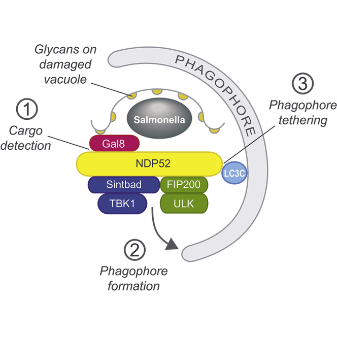
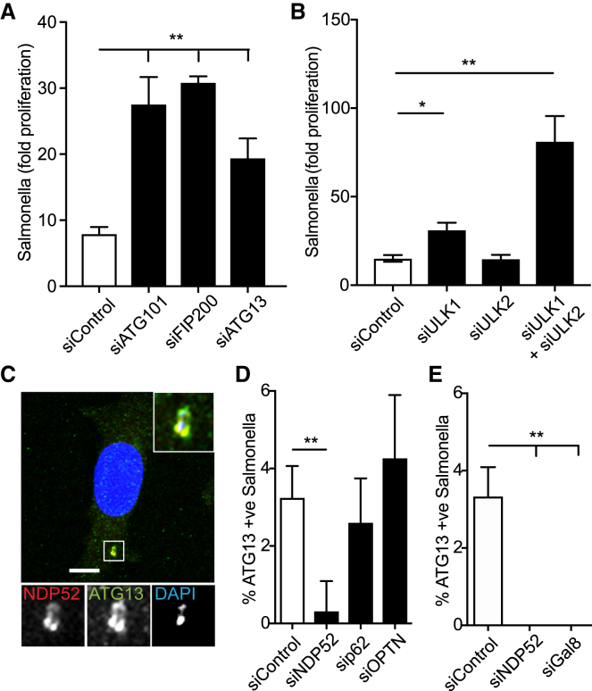
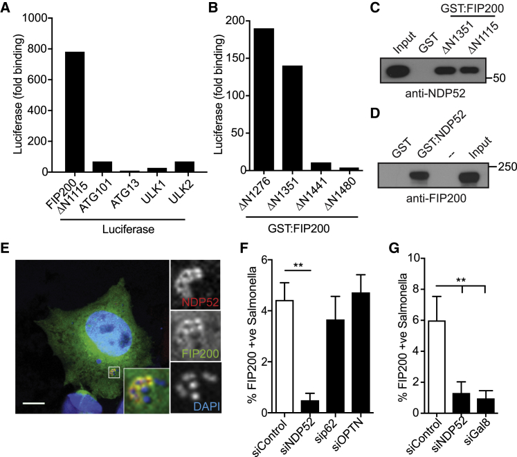
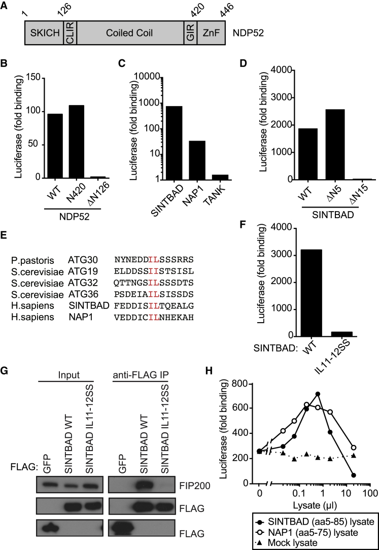
Xenophagy, a selective autophagy pathway that protects the cytosol against bacterial invasion, relies on cargo receptors that juxtapose bacteria and phagophore membranes. Whether phagophores are recruited from a constitutive pool or are generated de novo at prospective cargo remains unknown. Phagophore formation in situ would require recruitment of the upstream autophagy machinery to prospective cargo. Here, we show that, essential for anti-bacterial autophagy, the cargo receptor NDP52 forms a trimeric complex with FIP200 and SINTBAD/NAP1, which are subunits of the autophagy-initiating ULK and the TBK1 kinase complex, respectively. FIP200 and SINTBAD/NAP1 are each recruited independently to bacteria via NDP52, as revealed by selective point mutations in their respective binding sites, but only in their combined presence does xenophagy proceed. Such recruitment of the upstream autophagy machinery by NDP52 reveals how detection of cargo-associated "eat me" signals, induction of autophagy, and juxtaposition of cargo and phagophores are integrated in higher eukaryotes.

Keywords: FIP200; NDP52; Salmonella enterica; TBK1; ULK; cargo receptor; galectin-8; selective autophagy; xenophagy.


Figures

None
Graphical abstract
Figure 1
Figure 1
NDP52-Dependent Recruitment of the ULK Complex to S. Typhimurium Is Required for Restriction of Bacterial Proliferation (A) HeLa cells transfected with the indicated siRNAs were infected with S. Typhimurium. Colony-forming unit assay was used to assess the proliferation of bacteria over time. Data are depicted as fold proliferation of bacteria at 8 h versus 2 h post infection (p.i.). Mean ± SEM of at least three independent experiments is shown. (B) HeLa cells transfected with the indicated siRNAs were infected with S. Typhimurium. Colony-forming unit assay was used to assess the proliferation of bacteria over time. Data are depicted as fold proliferation of bacteria at 8 h versus 2 h post infection (p.i.). Mean ± SEM of at least three independent experiments is shown. (C–E) HeLa cells infected with S. Typhimurium were fixed at 1 h p.i. and stained for endogenous NDP52 and ATG13 (C) or ATG13 alone (D and E). (C) A representative confocal micrograph is depicted. DAPI signal in inset represents bacteria. (D) HeLa cells transfected with the indicated siRNAs were infected with S. Typhimurium. The frequency of ATG13-positive bacteria in cells transfected with the indicated siRNAs was enumerated on a wide-field microscope by eye. (E) The frequency of ATG13-positive bacteria in cells transfected with the indicated siRNAs was enumerated on a wide-field microscope by eye. Mean ± SD from two independent experiments. p < 0.05; ∗∗p < 0.01; one-way ANOVA with Dunnett’s multiple comparisons test. Scale bar, 10 μm.
Figure 2
Figure 2
NDP52 Binds FIP200 and Recruits It to Cytosolic S. Typhimurium (A) The indicated Luciferase-tagged constructs were expressed in HEK293ET cells and assessed for their ability to bind glutathione S-transferase (GST):NDP52 purified from E. coli in a LUMIER assay. (B) The indicated C-terminal fragments of GST:FIP200 were expressed in E. coli and assessed for their ability to bind Luciferase:NDP52 from HEK293ET cell lysate by LUMIER assay. (C and D) Bacterially expressed (C) FIP200 fragments or (D) NDP52 were assessed for their ability to bind endogenous NDP52 or FIP200, respectively, from HeLa cell lysates. Input = 10%. (E–G) Either (E) HeLa cells expressing FIP200ΔN1115:GFP alone or (F and G) transfected with the indicated siRNAs were infected with S. Typhimurium, fixed at 1 h p.i., and stained for endogenous NDP52. (E) A representative confocal micrograph is depicted. (F) The frequency of FIP200-positive bacteria was enumerated on a wide-field microscope by eye. Data in (A) and (B) are expressed as fold binding relative to GST alone. Data are from a single experiment representative of at least two (A–D) or mean ± SD of 2 independent experiments (F and G). ∗∗p < 0.01; one-way ANOVA with Dunnett’s multiple comparisons test. Scale bar, 10 μm.
Figure 3
Figure 3
FIP200 Binds the TBK1 Adaptor Proteins SINTBAD and NAP1 (A) Domain structure of NDP52. CLIR, LC3C-specific LC3-interacting region; GIR, galectin-8-interacting region; ZnF, zinc fingers. (B) Bacterially expressed GST-FIP200ΔN1115 was assessed for binding to the indicated Luciferase-tagged proteins from HEK293ET cell lysates by LUMIER assay. Data are from a single experiment representative of at least two independent experiments. (C) Bacterially expressed GST-FIP200ΔN1115 was assessed for binding to the indicated Luciferase-tagged proteins from HEK293ET cell lysates by LUMIER assay. Data are from a single experiment representative of at least two independent experiments. (D) Bacterially expressed GST-FIP200ΔN1115 was assessed for binding to the indicated Luciferase-tagged proteins from HEK293ET cell lysates by LUMIER assay. Data are from a single experiment representative of at least two independent experiments. (E) Alignment of the di-aliphatic amino-acid-containing regions of the indicated yeast cargo receptors and human SINTBAD and NAP1. Conserved residues within the motif are in red. (F) Bacterially expressed GST-FIP200ΔN1115 was assessed for binding to the indicated Luciferase-tagged proteins from HEK293ET cell lysates by LUMIER assay. Data are from a single experiment representative of at least two independent experiments. (G) The indicated FLAG-tagged constructs were expressed in HeLa cells, immunoprecipitated, and assessed for their ability to bind endogenous FIP200. Data are from a single experiment representative of three. (H) GST-FIP200ΔN1115 was assessed for its ability to bind Luciferase:NDP52 in the presence or absence of the indicated amounts of bacterial lysates containing SINTBADaa5-85, NAP1aa5-75, or mock by LUMIER assay. Data are expressed as fold binding relative to GST alone and are from a single experiment representative of three.
Figure 4
Figure 4
Identification of the Binding Sites for SINTBAD/NAP1 and FIP200 in the NDP52 SKICH Domain (A) GST:FIP200ΔN1115 was assessed for its ability to bind the indicated Luciferase-tagged proteins from HEK293ET cell lysates by LUMIER assay. Data are expressed as fold binding relative to GST alone and are from a single experiment representative of at least two. (B) Crystal structure of NDP52 SKICH domain with proposed binding residues highlighted. (C) The indicated FLAG-tagged proteins were assessed for their ability to bind the indicated Luciferase:NDP52 proteins from HEK293ET cell lysates by LUMIER assay. Data are from a single experiment representative of three independent experiments. (D) GST:FIP200ΔN1115 was assessed for its ability to bind the indicated Luciferase-tagged proteins from HEK293ET cell lysates by LUMIER assay. Data are expressed as fold binding relative to GST alone and are from a single experiment representative of at least two. (E) Schematic of NDP52 SKICH domain crystal structure with residues required for binding to FIP200 in red. (F) GST:FIP200ΔN1115 was assessed for its ability to bind the indicated Luciferase-tagged proteins from HEK293ET cell lysates by LUMIER assay. Data are expressed as fold binding relative to GST alone and are from a single experiment representative of at least two.
Figure 5
Figure 5
Identification of the Binding Site for SINTBAD/NAP1 in FIP200 (A and B) Bacterially expressed (A) GST:SINTBADaa5-85 or (B) GST:NDP52 were assessed for their ability to bind the indicated FIP200ΔN1115:Luciferase proteins from HEK293ET cell lysates by LUMIER assay. (C) GST:SINTBADaa5–85 or GST:NAP1aa5–75 was assessed for their ability to bind the indicated FIP200ΔN1115-Luciferase proteins from HEK293ET cell lysates by LUMIER assay. Data are expressed as binding relative to GST only versus FIP200ΔN1115 wild-type (WT):Luciferase and are from a single experiment representative of (A and B) two or (C) three.
Figure 6
Figure 6
Xenophagy Requires Simultaneous Recruitment of the ULK and SINTBAD/NAP1 Complexes to Cytosolic Bacteria via NDP52 (A–D) HeLa cells stably expressing (A) FIP200ΔN1115:GFP, (B) GFP:SINTBAD, (C) GFP:WIPI1, or (D) GFP:LC3B either alone (−) or together with the indicated NDP52 alleles were transfected with either control or NDP52 siRNA and infected with mCherry-S. Typhimurium for 1 h. Cells were fixed, and the recruitment of GFP-tagged proteins to S. Typhimurium was enumerated on a wide-field microscope by eye. Data are mean ± SEM of three independent experiments (A and B), mean ± SD of 2 independent experiments (C), or representative of at least two independent experiments (D). p < 0.05; ∗∗p < 0.01; one-way ANOVA with Dunnett’s multiple comparisons test.
Figure 7
Figure 7
Recruitment of the ULK Complex to Damaged SCV Membranes Initiates Phagophore Formation In Situ (A and B) HeLa cells stably expressing FIP200ΔN1115:GFP were infected with BFP-S. Typhimurium for 1 h, fixed, and stained with (A) anti-galectin-8 and anti-NDP52 antibodies or (B) anti-NDP52 and anti-WIPI2 antibodies. Images were acquired by super-resolution microscopy and are shown as maximum intensity projections. Yellow arrowheads denote WIPI2-positive omegasome-like structures. Scale bars, 1 μm.

Comment in

References

    1. Benjamin J.L., Sumpter R., Jr., Levine B., Hooper L.V. Intestinal epithelial autophagy is essential for host defense against invasive bacteria. Cell Host Microbe. 2013;13:723–734. - PMC - PubMed
    1. Bouwmeester T., Bauch A., Ruffner H., Angrand P.-O., Bergamini G., Croughton K., Cruciat C., Eberhard D., Gagneur J., Ghidelli S. A physical and functional map of the human TNF-α/NF-κ B signal transduction pathway. Nat. Cell Biol. 2004;6:97–105. - PubMed
    1. Boyle K.B., Randow F. The role of ‘eat-me’ signals and autophagy cargo receptors in innate immunity. Curr. Opin. Microbiol. 2013;16:339–348. - PubMed
    1. Deretic V., Saitoh T., Akira S. Autophagy in infection, inflammation and immunity. Nat. Rev. Immunol. 2013;13:722–737. - PMC - PubMed
    1. Dikic I., Elazar Z. Mechanism and medical implications of mammalian autophagy. Nat. Rev. Mol. Cell Biol. 2018;19:349–364. - PubMed

Publication types

MeSH terms

LinkOut - more resources