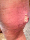
Large Undifferentiated Pleomorphic Sarcoma of the Posterior Thigh
- PMID: 30853708
- PMCID: PMC6421979
- DOI: 10.12659/AJCR.914079
Large Undifferentiated Pleomorphic Sarcoma of the Posterior Thigh
Abstract
BACKGROUND Sarcomas account for less than 1% of all cancers. Undifferentiated Pleomorphic Sarcoma, formerly called Malignant Fibrous Histiocytoma, is a rare subtype identified by a lack specific immunohistochemical markers for a specific lineage of differentiation. These soft tissue tumors are aggressive and rapidly enlarge. Risk for metastasis increases almost linearly as the tumor increases in size, emphasizing the importance of early detection, treatment, and post-resection monitoring. CASE REPORT This article reports a case of a large undifferentiated pleomorphic sarcoma of the posterior thigh in a 62-year-old female. Given the patient's history of thrombotic thrombocytopenic purpure, her initial mass was thought to be a hematoma following a hernia repair surgery. After diagnosis of undifferentiated pleomorphic sarcoma, she underwent radical excision revealing a 24×9.5×7cm lesion - one of the largest reported in the literature. CONCLUSIONS Sarcomas are very rare soft tissue neoplasms, but they should not be excluded in a physician's differentials when a patient presents with an enlarging soft tissue mass. Because sarcomas enlarge rapidly, delay in evaluation and management should be avoided and these patients should be quickly referred to a center specializing in sarcoma treatment. Magnetic Resonance Imaging (MRI) is the recommended initial imaging for all soft tissue masses of the extremities, trunk, and head and neck while Computed Tomography (CT) is the recommended imaging choice for retroperitoneal and visceral masses. After successful surgical excision with clean margins, patients should undergo serial monitoring by CT or MRI for surveillance of recurrence or late pulmonary metastases.
Conflict of interest statement
Figures
References
-
- Doyle LA. Sarcoma classification: an update based on the 2013 World Health Organization Classification of Tumors of Soft Tissue and Bone. Cancer. 2014;120(12):1763–74. - PubMed
-
- Demetri GD, Antonia S, Benjamin R, et al. Soft tissue sarcoma. J Natl Compr Canc Netw. 2010;8(6):630–74. - PubMed
-
- Morris CD. Malignant fibrous histiocytoma. Liddy Shriver Sarcoma Initiative. 2005. http://sarcomahelp.org/mfh.html#tpm1_1.
-
- Chen KH, Chou TM, Shieh SJ. Management of extremity malignant fibrous histiocytoma: A 10-year experience. Formosan Journal of Surgery. 2015;48(1):1–9.
Publication types
MeSH terms
LinkOut - more resources
Full Text Sources


