Prenatal Protein Malnutrition Produces Resistance to Distraction Similar to Noradrenergic Deafferentation of the Prelimbic Cortex in a Sustained Attention Task
- PMID: 30853881
- PMCID: PMC6396814
- DOI: 10.3389/fnins.2019.00123
Prenatal Protein Malnutrition Produces Resistance to Distraction Similar to Noradrenergic Deafferentation of the Prelimbic Cortex in a Sustained Attention Task
Abstract
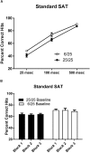
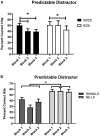
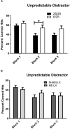
Exposure to malnutrition early in development increases likelihood of neuropsychiatric disorders, affective processing disorders, and attentional problems later in life. Many of these impairments are hypothesized to arise from impaired development of the prefrontal cortex. The current experiments examine the impact of prenatal malnutrition on the noradrenergic and cholinergic axons in the prefrontal cortex to determine if these changes contribute to the attentional deficits seen in prenatal protein malnourished rats (6% casein vs. 25% casein). Because prenatally malnourished animals had significant decreases in noradrenergic fibers in the prelimbic cortex with spared innervation in the anterior cingulate cortex and showed no changes in acetylcholine innervation of the prefrontal cortex, we compared deficits produced by malnutrition to those produced in adult rats by noradrenergic lesions of the prelimbic cortex. All animals were able to perform the baseline sustained attention task accurately. However, with the addition of visual distractors to the sustained attention task, animals that were prenatally malnourished and those that were noradrenergically lesioned showed cognitive rigidity, i.e., were less distractible than control animals. All groups showed similar changes in behavior when exposed to withholding reinforcement, suggesting specific attentional impairments rather than global difficulties in understanding response rules, bottom-up perceptual problems, or cognitive impairments secondary to dysfunction in sensitivity to reinforcement contingencies. These data suggest that prenatal protein malnutrition leads to deficits in noradrenergic innervation of the prelimbic cortex associated with cognitive rigidity.
Keywords: cognition; distractibility; norepinephrine; prefrontal cortex; rat model of malnutrition.
Figures


Similar articles
-
The neural basis of attentional alterations in prenatally protein malnourished rats.Cereb Cortex. 2021 Jan 1;31(1):497-512. doi: 10.1093/cercor/bhaa239. Cereb Cortex. 2021. PMID: 33099611 Free PMC article.
-
Prenatal malnutrition leads to deficits in attentional set shifting and decreases metabolic activity in prefrontal subregions that control executive function.Dev Neurosci. 2014;36(6):532-41. doi: 10.1159/000366057. Epub 2014 Oct 22. Dev Neurosci. 2014. PMID: 25342495
-
Noradrenergic, but not cholinergic, deafferentation of prefrontal cortex impairs attentional set-shifting.Neuroscience. 2008 Apr 22;153(1):63-71. doi: 10.1016/j.neuroscience.2008.01.064. Epub 2008 Feb 19. Neuroscience. 2008. PMID: 18355972 Free PMC article.
-
Prefrontal executive and cognitive functions in rodents: neural and neurochemical substrates.Neurosci Biobehav Rev. 2004 Nov;28(7):771-84. doi: 10.1016/j.neubiorev.2004.09.006. Neurosci Biobehav Rev. 2004. PMID: 15555683 Review.
-
Differential cognitive actions of norepinephrine a2 and a1 receptor signaling in the prefrontal cortex.Brain Res. 2016 Jun 15;1641(Pt B):189-96. doi: 10.1016/j.brainres.2015.11.024. Epub 2015 Nov 22. Brain Res. 2016. PMID: 26592951 Free PMC article. Review.
Cited by
-
Impact of Early Childhood Malnutrition on Adult Brain Function: An Evoked-Related Potentials Study.Front Hum Neurosci. 2022 Jul 1;16:884251. doi: 10.3389/fnhum.2022.884251. eCollection 2022. Front Hum Neurosci. 2022. PMID: 35845242 Free PMC article.
-
Prenatal protein malnutrition decreases neuron numbers in the parahippocampal region but not prefrontal cortex in adult rats.Nutr Neurosci. 2025 Mar;28(3):333-346. doi: 10.1080/1028415X.2024.2371256. Epub 2024 Aug 1. Nutr Neurosci. 2025. PMID: 39088448
-
Prenatal Protein Malnutrition Leads to Hemispheric Differences in the Extracellular Concentrations of Norepinephrine, Dopamine and Serotonin in the Medial Prefrontal Cortex of Adult Rats.Front Neurosci. 2019 Mar 5;13:136. doi: 10.3389/fnins.2019.00136. eCollection 2019. Front Neurosci. 2019. PMID: 30890908 Free PMC article.
-
The neural basis of attentional alterations in prenatally protein malnourished rats.Cereb Cortex. 2021 Jan 1;31(1):497-512. doi: 10.1093/cercor/bhaa239. Cereb Cortex. 2021. PMID: 33099611 Free PMC article.
-
The Central Noradrenergic System in Neurodevelopmental Disorders: Merging Experimental and Clinical Evidence.Int J Mol Sci. 2023 Mar 18;24(6):5805. doi: 10.3390/ijms24065805. Int J Mol Sci. 2023. PMID: 36982879 Free PMC article. Review.
References
-
- Berridge C. W., Shumsky J. S., Andrzejewski M. E., McGaughy J. A., Spencer R. C., Devilbiss D. M., et al. (2012). Differential sensitivity to psychostimulants across prefrontal cognitive tasks: differential involvement of noradrenergic alpha(1) - and alpha(2)-receptors. Biol. Psychiatry 71 467–473. 10.1016/j.biopsych.2011.07.022 - DOI - PMC - PubMed
Grants and funding
LinkOut - more resources
Full Text Sources

