. 2019 Feb 22:13:137.
doi: 10.3389/fnins.2019.00137. eCollection 2019.

Cell Type-Specific Human APP Transgene Expression by Hippocampal Interneurons in the Tg2576 Mouse Model of Alzheimer's Disease

Affiliations

Cell Type-Specific Human APP Transgene Expression by Hippocampal Interneurons in the Tg2576 Mouse Model of Alzheimer's Disease

Corinna Höfling et al. Front Neurosci. .

Abstract

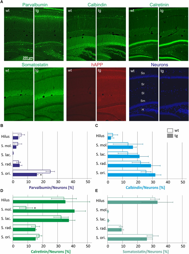
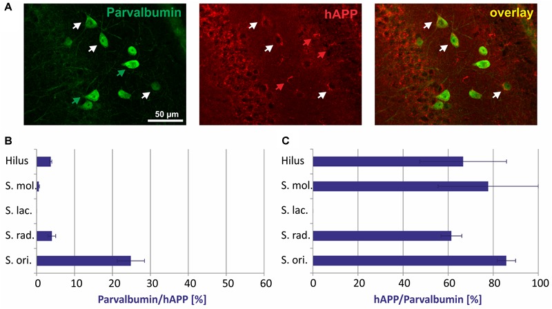
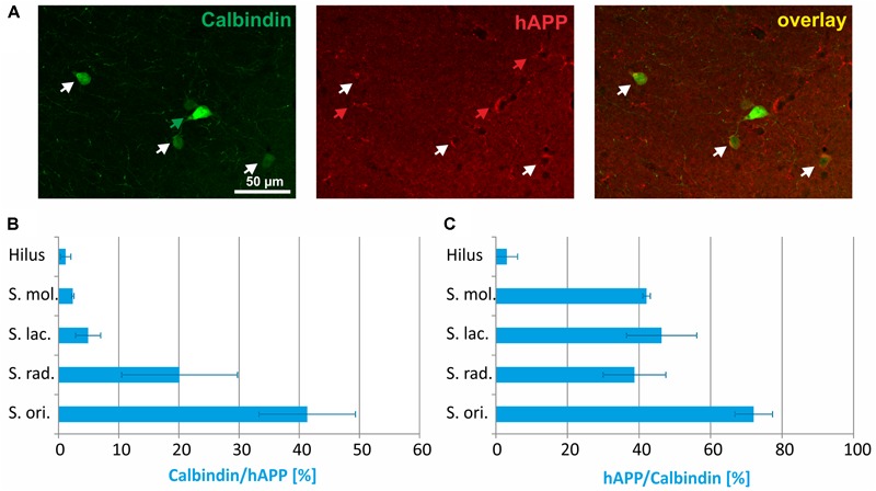
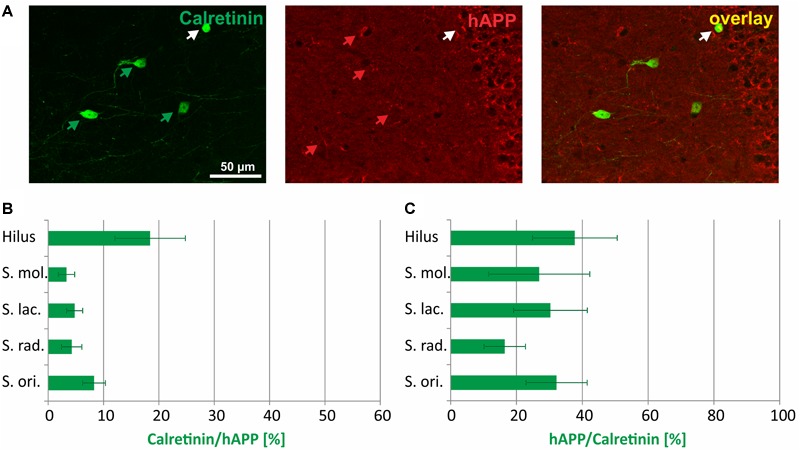
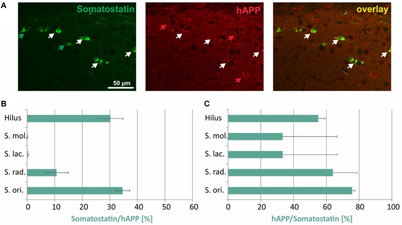
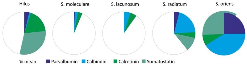
Amyloid precursor protein (APP) transgenic animal models of Alzheimer's disease have become versatile tools for basic and translational research. However, there is great heterogeneity of histological, biochemical, and functional data between transgenic mouse lines, which might be due to different transgene expression patterns. Here, the expression of human APP (hAPP) by GABAergic hippocampal interneurons immunoreactive for the calcium binding proteins parvalbumin, calbindin, calretinin, and for the peptide hormone somatostatin was analyzed in Tg2576 mice by double immunofluorescent microscopy. Overall, there was no GABAergic interneuron subpopulation that did not express the transgene. On the other hand, in no case all neurons of such a subpopulation expressed hAPP. In dentate gyrus molecular layer and in stratum lacunosum moleculare less than 10% of hAPP-positive interneurons co-express any of these interneuron markers, whereas in stratum oriens hAPP-expressing neurons frequently co-express these interneuron markers to different proportions. We conclude that these neurons differentially contribute to deficits in young Tg2576 mice before the onset of Abeta plaque pathology. The detailed analysis of distinct brain region and neuron type-specific APP transgene expression patterns is indispensable to understand particular pathological features and mouse line-specific differences in neuronal and systemic functions.

Keywords: Alzheimer’s disease; amyloid precursor protein; animal model; calcium-binding proteins; hippocampus; interneuron.


Figures

FIGURE 1
FIGURE 1
Quantification of interneuron numbers in hippocampal layers of wild type (wt) and hAPP-transgenic Tg2576 mice (tg). In panel (A) typical staining patterns of parvalbumin-, calbindin-, calretinin-, somatostatin-, and hAPP-immunoreactive neurons are depicted in wt and tg mouse hippocampus. Note the similar labeling intensity in wt and tg brain sections for all antigens except hAPP. The hippocampal layers analyzed are indicated in the wt pan-neuronal labeling image (Neurons). In panels B–E, the proportions of parvalbumin-, calbindin-, calretinin-, and somatostatin-positive neurons in hippocampal layers of wt and tg mice are quantified. Total neuronal numbers in each layer (hilus 69.8 ± 2.8; S. mol 48.2 ± 6.7; S. lac. 68.5 ± 8.5; S. rad. 407.2 ± 49.0; S. ori. 269.5 ± 13.0) were set to 100%. Note the similar interneuron proportions in wt and tg hippocampus for most data analyzed. significantly different from wild type (p < 0.05; unpaired t test).
FIGURE 2
FIGURE 2
Quantitative analyses of the co-localization between parvalbumin-immunoreactive neurons and hAPP-expressing interneurons in hippocampus of Tg2576 mice. Typical examples of double labelings in stratum oriens are shown in panel (A). White arrows point to double-labeled neurons, green, and red arrows to neurons that are only labeled by green (parvalbumin) and red (hAPP) fluorescence, respectively. Quantitative analysis revealed that of all hAPP transgene-expressing interneurons 0% (in stratum lacunosum moleculare) to 25% (in stratum oriens) were parvalbumin-immunoreactive (B). Of all parvalbumin-expressing neurons, 60 to 80% expressed the hAPP transgene in hilus of DG, stratum moleculare, stratum radiatum, and stratum oriens (C).
FIGURE 3
FIGURE 3
Quantitative analyses of the co-localization between calbindin-immunoreactive neurons and hAPP-expressing interneurons in hippocampus of Tg2576 mice. Typical examples of double labelings in stratum radiatum are shown in panel (A). White arrows point to double-labeled neurons, green, and red arrows to neurons that are only labeled by green (calbindin) and red (hAPP) fluorescence, respectively. Quantitative analysis revealed that of all hAPP transgene-expressing interneurons 2% (in DG hilus) to 42% (in stratum oriens) were calbindin-immunoreactive (B). Of all calbindin-expressing neurons, 5 to 70% expressed the hAPP transgene (C).
FIGURE 4
FIGURE 4
Quantitative analyses of the co-localization between calretinin-immunoreactive neurons and hAPP-expressing interneurons in hippocampus of Tg2576 mice. Typical examples of double labelings in stratum radiatum are shown in panel (A). White arrows point to double-labeled neurons, green, and red arrows to neurons that are only labeled by green (calretinin) and red (hAPP) fluorescence, respectively. Quantitative analysis revealed that of all hAPP transgene-expressing interneurons 4% (in stratum moleculare) to 18% (in DG hilus) were calretinin-immunoreactive (B). Of all calretinin-expressing neurons, 18 to 38% expressed the hAPP transgene in defined hippocampal layers (C).
FIGURE 5
FIGURE 5
Quantitative analyses of the co-localization between somatostatin-immunoreactive neurons and hAPP-expressing interneurons in hippocampus of Tg2576 mice. Typical examples of double labelings in stratum oriens are shown in panel (A). White arrows point to double-labeled neurons, green, and red arrows to neurons that are only labeled by green (somatostatin) and red (hAPP) fluorescence, respectively. Quantitative analysis revealed that of all hAPP transgene-expressing interneurons 1 to 2% in stratum moleculare and stratum lacunosum moleculare to approximately 30% (in DG hilus and stratum oriens) were somatostatin- immunoreactive (B). Of all somatostatin-expressing neurons, 30 to 76% expressed the hAPP transgene in all hippocampal layers (C).
FIGURE 6
FIGURE 6
In this Figure, the proportion of hAPP-positive hippocampal interneurons co-expressing parvalbumin, calbindin, calretinin, and somatostatin is summed up in individual hippocampal layers. In stratum moleculare and stratum lacunosum moleculare less than 10% of all hAPP-expressing neurons are immunoreactive for the four interneuron markers analyzed, whereas in stratum radiatum and DG hilus 40 to 50%, and in stratum oriens all hAPP-immunoreactive neurons are positive for these interneuron markers. Additionally, the contribution of individual interneuron types to the hAPP-expressing population varies considerably in hippocampal layers as indicated by the respective color.

Similar articles

Cited by

References

    1. Allen G., Barnard H., McColl R., Hester A. L., Fields J. A., Weiner M. F., et al. (2007). Reduced hippocampal functional connectivity in Alzheimer disease. Arch. Neurol. 64 1482–1487. 10.1001/archneur.64.10.1482 - DOI - PubMed
    1. Ameen-Ali K. E., Wharton S. B., Simpson J. E., Heath P. R., Sharp P., Berwick J. (2017). Review: neuropathology and behavioural features of transgenic murine models of Alzheimer’s disease. Neuropathol. Appl. Neurobiol. 43 553–570. 10.1111/nan.12440 - DOI - PubMed
    1. Baglietto-Vargas D., Moreno-Gonzalez I., Sanchez-Varo R., Jimenez S., Trujillo-Estrada L., Sanchez-Mejias E., et al. (2010). Calretinin interneurons are early targets of extracellular amyloid-beta pathology in PS1/AbetaPP Alzheimer mice hippocampus. J. Alzheimers Dis. 21 119–132. 10.3233/JAD-2010-100066 - DOI - PubMed
    1. Barthel H., Sabri O. (2017). Clinical use and utility of amyloid imaging. J. Nucl. Med. 58 1711–1717. 10.2967/jnumed.116.185017 - DOI - PubMed
    1. Brady D. R., Mufson E. J. (1997). Parvalbumin-immunoreactive neurons in the hippocampal formation of Alzheimer’s diseased brain. Neuroscience 80 1113–1125. 10.1016/S0306-4522(97)00068-7 - DOI - PubMed

LinkOut - more resources