Review
. 2019 Feb 3:2019:5268342.
doi: 10.1155/2019/5268342. eCollection 2019.

Resin-Dentin Bonding Interface: Mechanisms of Degradation and Strategies for Stabilization of the Hybrid Layer

Affiliations
Review

Resin-Dentin Bonding Interface: Mechanisms of Degradation and Strategies for Stabilization of the Hybrid Layer

D E Betancourt et al. Int J Biomater. .

Abstract

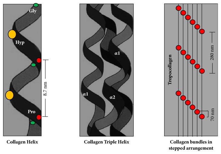
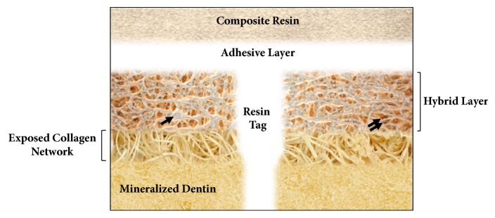
Several studies have shown that the dentin-resin interface is unstable due to poor infiltration of resin monomers into the demineralized dentin matrix. This phenomenon is related to the incomplete infiltration of the adhesive system into the network of exposed collagen fibrils, mainly due to the difficulty of displacement and subsequent replacement of trapped water between interfibrillar spaces, avoiding adequate hybridization within the network of collagen fibrils. Thus, unprotected fibrils are exposed to undergo denaturation and are susceptible to cyclic fatigue rupture after being subjected to repetitive loads during function. The aqueous inclusions within the hybrid layer serve as a functional medium for the hydrolysis of the resin matrix, giving rise to the activity of esterases and collagenolytic enzymes, such as matrix metalloproteinases, which play a fundamental role in the degradation process of the hybrid layer. Achieving better interdiffusion of the adhesive system in the network of collagen fibrils and the substrate stability in the hybrid layer through different strategies are key events for the interfacial microstructure to adequately function. Hence, it is important to review the factors related to the mechanisms of degradation and stabilization of the hybrid layer to support the implementation of new materials and techniques in the future. The enzymatic degradation of collagen matrix, together with resin leaching, has led to seeking strategies that inhibit the endogenous proteases, cross-linking the denudated collagen fibrils and improving the adhesive penetration removing water from the interface. Some of dentin treatments have yielded promising results and require more research to be validated. A longer durability of adhesive restorations could resolve a variety of clinical problems, such as microleakage, recurrent caries, postoperative sensitivity, and restoration integrity.


Figures

Figure 1
Figure 1
Structure of collagen. Each collagen helix is called the α chain, which is a levorotatory strand with about 3 residues per turn of glycine (Gly), proline (Pro), and hydroxyproline (Hyp) sequences. The quaternary structure of the collagen fibril is formed from the supercoiling of three α chains to form a triple dextrorotatory helix.
Figure 2
Figure 2
Location of SIBLING proteins in mineralized dentin. An immunocytochemistry analysis using one of two primary antibodies, polyclonal antidentin sialophosphoprotein (Abcam, Cambridge, MA, USA) which is specific to dentin sialoprotein (DSP) or polyclonal antidentinal matrix protein (DMP)-1 (Sigma-Aldrich, St Louis, MO, USA) and visualized using peroxidase (brown color in (a) and (c)) or Alexa Fluor 594 (red color in (b) and (d)) coupled streptavidin under fluorescence microscopy revealed the localization patterns of DSP and DMP-1. DSP and DMP-1 are strongly expressed in the odontoblastic layer (arrow) and with less intensity in the predentin area (double arrow) and in the dentin mineralization front (head arrow). (a) and (b) immunopositive staining for DSP in the mineralized dentin. (c) and (d) immunolocalization of DMP-1. These non-collagenous proteins interact with collagen fibrils and control initiation and growth of apatite crystals on dentinal matrix. Mineralized dentin counterstained with hematoxylin (a and c) and Hoechst blue 33342 (b and d). Bar corresponds to 100 μm. Courtesy of Baldion et al. [29].
Figure 3
Figure 3
Basic structure of the MMP. MMPs consist of four main domains: a signal peptide that directs the secretion of the protein to the outside of the cell, a propeptide region that keeps the enzyme inactive until it is proteolytically cleaved, a catalytic domain that contains zinc and calcium and to which the cysteine of the propeptide region binds to keep it inactive (cysteine switch), and a hemopexin-like domain that mediates substrate specificity and interactions with endogenous inhibitors. MMP-2 and MMP-9 possess a fibronectin-type domain for matrix binding.
Figure 4
Figure 4
Resin-dentin interface with 150 days of aging by SEM. (a) Arrows show voids in the deepest portion of hybrid layer. (b) Degradation of collagen fibrils is evidenced (arrows) in the adhesive interface. (c) Loss of collagen in the intertubular dentin around the resin tags (arrows). (d) Degradation of the bonding interface with formation of water channels (arrows) and hydrolytic degradation of the resin adhesive. Bonded interfaces were created with Adper Single Bond 2 (3M ESPE, St. Paul, MN, USA). (SEM) scanning electron microscopy, (ITD) intertubular dentin, (T) dentin tubule, (DC) degraded collagen, (RT) resin tags, (HL) hybrid layer, (AL) adhesive layer. Bar corresponding to 10µm. Courtesy of Betancourt DE and Baldion PA with permission.
Figure 5
Figure 5
Schematic illustration of the instability of the hybrid layer. Apatite crystals are removed from dental hard tissues with acid conditioning and should be replaced by resin monomers. Note the incomplete infiltration of the adhesive in the collagen matrix that remains with interfibrillar spaces saturated with water (arrow). In addition, the possibility of discrepancy between the depth of infiltration of the adhesive and that of conditioned dentin leaving collagen exposed without hybridizing. The resin tags partially seal the dentinal tubules and decrease the permeability of the dentin. The resin monomers do not penetrate homogenously into the collagenous network (double arrow) [58] (with permission).

References

    1. Van Landuyt K. L., Snauwaert J., De Munck J., et al. Systematic review of the chemical composition of contemporary dental adhesives. Biomaterials. 2007;28(26):3757–3785. doi: 10.1016/j.biomaterials.2007.04.044. - DOI - PubMed
    1. Liu Y., Tjäderhane L., Breschi L., et al. Limitations in bonding to dentin and experimental strategies to prevent bond degradation. Journal of Dental Research. 2011;90(8):953–968. doi: 10.1177/0022034510391799. - DOI - PMC - PubMed
    1. Nakabayashi N., Nakamura M., Yasuda N. Hybrid Layer as a Dentin-Bonding Mechanism. Journal of Esthetic and Restorative Dentistry. 1991;3(4):133–138. doi: 10.1111/j.1708-8240.1991.tb00985.x. - DOI - PubMed
    1. Pashley D. H., Tay F. R., Breschi L., et al. State of the art etch-and-rinse adhesives. Dental Materials. 2011;27(1):1–16. doi: 10.1016/j.dental.2010.10.016. - DOI - PMC - PubMed
    1. Mazzoni A., Scaffa P., Carrilho M., et al. Effects of etch-and-rinse and self-etch adhesives on dentin MMP-2 and MMP-9. Journal of Dental Research. 2013;92(1):82–86. doi: 10.1177/0022034512467034. - DOI - PMC - PubMed

LinkOut - more resources