. 2019 Mar;17(3):2639-2646.
doi: 10.3892/ol.2019.9881. Epub 2019 Jan 2.

Regulation of OLC1 protein expression by the anaphase-promoting complex

Affiliations

Regulation of OLC1 protein expression by the anaphase-promoting complex

Xiaojing Zhang et al. Oncol Lett. 2019 Mar.

Abstract

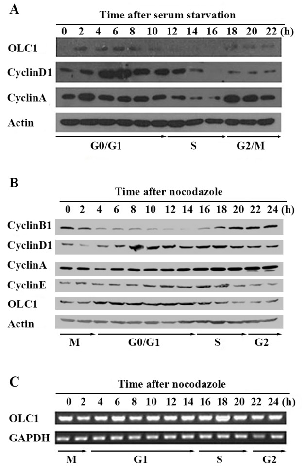
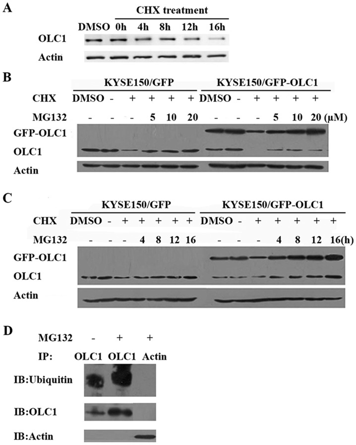
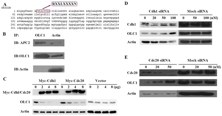
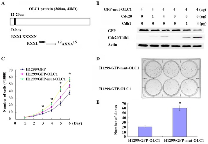
Overexpressed in lung cancer 1 (OLC1) is a potential oncogene overexpressed in human lung cancer and in other types of malignant tumor. The elevated expression of OLC1 contributes to tumor genesis and progression. However, the mechanisms regulating the expression of OLC1 remain unclear. In the present study, using lung and esophageal cancer cell lines, it was demonstrated that OLC1 was a short-lived, cell cycle-dependent protein regulated through the anaphase-promoting complex/cyclosome (APC/c)-ubiquitin pathway by directly interacting with the APC2 subunit. Through the action of two co activator proteins, cadherin 1 (Cdh1) and cell-division cycle protein 20 (Cdc20), the OLC1 protein was ubiquitinated and degraded. Following treatment with a proteasome inhibitor, OLC1 protein levels were elevated. Inversely, the upregulation of Cdh1 and Cdc20 facilitated OLC1 degradation. By inducing point mutations of the assumed degradation motif of OLC1, it was revealed that an intact destruction (D)-box was necessary. As expected, the D-box-mutated OLC1 exhibited a higher capacity for promoting cell growth and clone formation. Collectively, these findings indicate that the expression of the candidate oncogene OLC1 is cell cycle-dependent and is regulated by an APC/c mediated ubiquitin-proteasome pathway.

Keywords: anaphase-promoting complex/cyclosome; cadherin 1; cell-division cycle protein 20; degradation; destruction box; overexpressed in lung cancer 1.


Figures

Figure 1.
Figure 1.
Expression of OLC1 throughout the cell cycle. (A) Using serum starvation, KYSE150/GFP cells were synchronized at the G0 phase. Following release, cells were collected every 2 for 24 h. The protein expression of OLC1, cyclin D1, cyclin A and actin was analyzed using western blot analysis. (B) Using 0.4 µg/ml nocodazole, KYSE150/GFP cells were synchronized at the mitotic phase. Following release, cells were collected every 2 for 24 h. The protein expression of OLC1, cyclin B1, cyclin D1, cyclin A, cyclin E and actin was analyzed using western blot analysis. (C) Using reverse transcription polymerase chain reaction, the mRNA expression of OLC1 and GAPDH were detected. OLC1, overexpressed in lung cancer 1.
Figure 2.
Figure 2.
OLC1 protein expression with CHX and MG132 treatment. (A) Following treatment with 100 µg/ml CHX, KYSE150/GFP cells were collected for western blot analysis of OLC1 protein expression at the indicated times. KYSE150/GFP and KYSE150/GFP-OLC1 cells were co-treated with CHX (100 µg/ml) and MG132 at (B) different concentrations (5, 10 and 20 µM MG132) or (C) for different lengths of time (4, 8, 12 and 16 h) with 20 µM MG132, with the DMSO, negative control and MG132-negative groups collected for analysis after 16 h of treatment. Results are representative of three independent experiments. (D) With (+) or without (−) treatment with 20 µM MG132, KYSE150/GFP-OLC1 cells were incubated for 16 h, collected and lysed for IP with an anti-OLC1 antibody. Ubiquitins were detected in the immunocomplex, and the presence of OLC1 was verified. IP with an anti-actin antibody was used as a negative control. OLC1, overexpressed in lung cancer 1; CHX, cycloheximide; IP, immunoprecipitation.
Figure 3.
Figure 3.
OLC1 protein is degraded by the APC/c. (A) Analysis of the OLC1 protein sequence indicated that it contains a destruction box at the site of amino acids 12–20, which may be recognized by APC/c. (B) To investigate whether OLC1 may bind directly to the components of the APC/c, KYSE150/GFP-OLC1 cells were collected, lysed and subjected to IP. The presence of actin, OLC1 and APC2 in the immunocomplex were verified. (C) Different concentrations (2, 4 and 8 µg) of Myc-Cdh1/Cdc20 expression or mock vectors were transiently transfected into H1299 cells for 48 h. Then OLC1, Myc-Cdh1 and Myc-Cdc20 protein expression were evaluated. Actin was included as a loading control. Different concentrations (20, 50 and 100 nM) of (D) Cdh1 or (E) Cdc20 or mock siRNA were transiently transfected into H1299 cells for 48 h. Then OLC1, Cdh1 and Cdc20 protein expressions were evaluated. Actin was included as a loading control. OLC1, overexpressed in lung cancer 1; APC/c, anaphase-promoting cyclosome complex; IP, immunoprecipitation; Cdh1, cadherin 1; Cdc20, cell-division cycle protein 20; siRNA, small interfering RNA.
Figure 4.
Figure 4.
Mutated D-box domains interfere with OLC1 protein degradation and accelerate cell growth and colony formation. (A) A schematic representation of the D-box degradation signal within OLC1. Numbers denote amino acid numbers where the D-box lies, and the conserved sequence motif is annotated. The mutation scheme for the OLC1 D-box is indicated. (B) H1299 cells were co-transfected with 4 µg OLC1 (D-box mutant) plasmids and different concentrations of human Myc-Cdh1/Cdc20 expression vectors for 48 h. OLC1, Cdh1 and Cdc20 protein expression were evaluated using western blot analysis. Actin was probed as a loading control. (C) H1299 cells were transfected with 4 µg pEGFP-N1, pEGFP-N1-OLC1 or pEGFP-N1-mut-OLC1. Mean values with standard deviations were calculated to produce a cell proliferation curve. The results were obtained in three independent experiments. *P<0.05 vs. wild-type OLC1. (D) A colony formation assay of H1299 cells was performed following transfections of a mutated or wild-typeOLC1 expression vector for 24 h and (E) the number of colonies were counted. The results were obtained from three independent experiments. *P<0.05 vs. control cells. OLC1, overexpressed in lung cancer 1; D-box, destruction box; Cdh1, cadherin 1; Cdc20, cell-division cycle protein 20.

Similar articles

Cited by

References

    1. Yuan J, Ma J, Zheng H, Shi T, Sun W, Zhang Q, Lin D, Zhang K, He J, Mao Y, et al. Overexpression of OLC1, cigarette smoke, and human lung tumorigenesis. J Natl Cancer Inst. 2008;100:1592–1605. doi: 10.1093/jnci/djn379. - DOI - PMC - PubMed
    1. Li X, Suo J, Shao S, Xue L, Chen W, Dong L, Shi J, Fu M, Lu N, Zhan Q, Tong T. Overexpression of OLC1 promotes tumorigenesis of human esophageal squamous cell carcinoma. PLoS One. 2014;9:e90958. doi: 10.1371/journal.pone.0090958. - DOI - PMC - PubMed
    1. Dimaano C, Jones CB, Hanono A, Curtiss M, Babst M. Ist1 regulates Vps4 localization and assembly. Mol Biol Cell. 2008;19:465–474. doi: 10.1091/mbc.e07-08-0747. - DOI - PMC - PubMed
    1. Rue SM, Mattei S, Saksena S, Emr SD. Novel Ist1-Did2 complex functions at a late step in multivesicular body sorting. Mol Biol Cell. 2008;19:475–484. doi: 10.1091/mbc.e07-07-0694. - DOI - PMC - PubMed
    1. Agromayor M, Carlton JG, Phelan JP, Matthews DR, Carlin LM, Ameer-Beg S, Bowers K, Martin-Serrano J. Essential role of hIST1 in cytokinesis. Mol Biol Cell. 2009;20:1374–1387. doi: 10.1091/mbc.e08-05-0474. - DOI - PMC - PubMed