. 2019 Feb 3:2019:8581743.
doi: 10.1155/2019/8581743. eCollection 2019.

Clinical Impact of EUS-Guided Fine Needle Biopsy Using a Novel Franseen Needle for Histological Assessment of Pancreatic Diseases

Affiliations

Clinical Impact of EUS-Guided Fine Needle Biopsy Using a Novel Franseen Needle for Histological Assessment of Pancreatic Diseases

Takuya Ishikawa et al. Can J Gastroenterol Hepatol. .

Abstract

Background and aims: Several studies have shown the benefits of endoscopic ultrasound-guided fine needle biopsy (EUS-FNB) using a Franseen needle for histological assessment. However, studies focusing on pancreatic diseases are limited and the safety of this method has not been well assessed. We aimed to assess the current status and issues of EUS-FNB in the diagnosis of pancreatic diseases.

Materials and methods: We retrospectively reviewed 87 consecutive EUS-FNB specimens using either a 22-gauge Franseen needle (Group A, N = 51) or a conventional 22-gauge fine-needle aspiration needle (Group B, N = 36) for pancreatic diseases, and the diagnostic accuracy and safety were compared. Final diagnoses were obtained based on surgical pathology or a minimum six-month clinical follow-up.

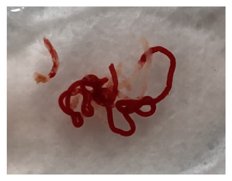
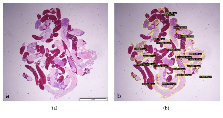
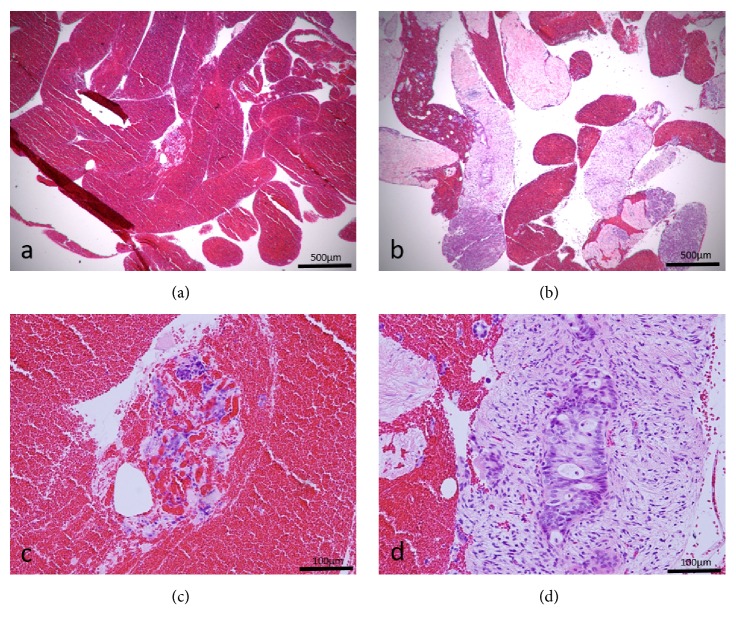
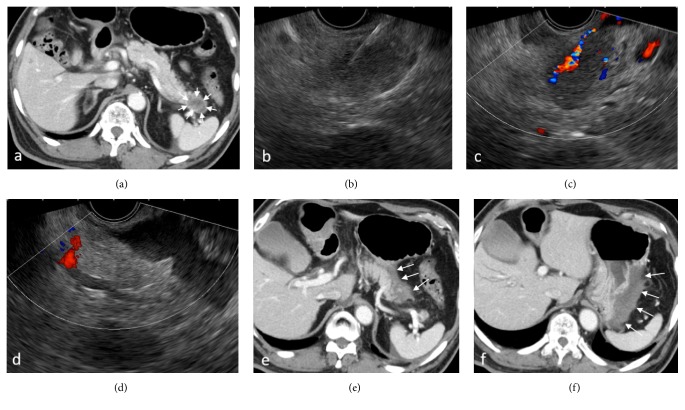
Results: Although the diagnostic accuracy for malignancy was 96.1% in Group A versus 88.9% in Group B, with no statistically significant difference (P = 0.19), the median sample area was significantly larger in Group A (4.07 versus 1.31mm2, P < 0.0001). There were no differences between the two needles in the locations from which the specimens were obtained. Adverse events occurred in one case (2%) in Group A (mild pancreatitis) and none in Group B with no statistical significance (P = 0.586). Although there was no case of bleeding defined as adverse events, 2 cases in Group A showed active bleeding during the procedure with increase in the echo-free space, which required CT scanning to rule out extravasation. Eventually, the bleeding stopped spontaneously.

Conclusions: Given its guaranteed ability to obtain core specimens and comparable safety, and although the risk of bleeding should be kept in mind, EUS-FNB using a Franseen needle is likely to become a standard procedure for obtaining pancreatic tissue in the near future.


Figures

Figure 1
Figure 1
Fresh specimen obtained with a 22-gauge Franseen needle. Fragments of whitish tissue can be detected macroscopically between the blood clots.
Figure 2
Figure 2
(a) Hematoxylin and eosin staining of a gross specimen obtained from the pancreas using a 22-gauge Franseen needle, viewed in a low-power field. (b) Measuring the area of the specimen, excluding the blood clots, using imaging software (CellSense).
Figure 3
Figure 3
Specimens obtained from the same pancreatic cancer lesion using both a conventional fine needle aspiration (FNA) needle (a, c) and a Franseen needle (b, d). The specimen obtained by the FNA needle shows many blood clots (a), whereas the specimen obtained by the Franseen needle shows good core tissues with low-power field magnification. (c) Scattered atypical cells can be identified in the blood clots with high-power field magnification, but they are insufficient for a cancer diagnosis. (d) A component of atypical cells with enlarged nuclei in the fibrous stroma is detected with high-power field magnification, consistent with ductal carcinoma of the pancreas.
Figure 4
Figure 4
A patient who developed bleeding with endoscopic ultrasound-guided fine needle biopsy (EUS-FNB) using a Franseen needle. (a) Contrast-enhanced Computed Tomography (CT) scan showing a 3-cm hypovascular mass in the pancreatic tail (arrow). (b) Insertion of the needle under EUS guidance. (c) Active bleeding from the needle tract right was noticed under color Doppler mode after the withdrawal of the needle. (d) Increased echo-free space between the pancreas and stomach was identified. ((e), (f)) Contrast-enhanced CT scan was performed immediately after EUS-FNB. Hyperdense fluid collection suggesting hematoma was observed between the pancreatic tail and the greater curvature of the stomach (arrow).

References

    1. Matsui M., Goto H., Niwa Y., Arisawa T., Hirooka Y., Hayakawa T. Preliminary results of fine needle aspiration biopsy histology in upper gastrointestinal submucosal tumors. Endoscopy. 1998;30(9):750–755. doi: 10.1055/s-2007-1001416. - DOI - PubMed
    1. Ishikawa T., Mohamed R., Heitman S. J., et al. Diagnostic yield of small histological cores obtained with a new EUS-guided fine needle biopsy system. Surgical Endoscopy. 2017;31(12):5143–5149. doi: 10.1007/s00464-017-5580-3. - DOI - PubMed
    1. Adler D., Muthusamy V. R., Ehrlich D., et al. A multicenter evaluation of a new EUS core biopsy needle: Experience in 200 patients. Endoscopic Ultrasound. 2018 doi: 10.4103/eus.eus_53_17. - DOI - PMC - PubMed
    1. Mukai S., Itoi T., Yamaguchi H., et al. A retrospective histological comparison of EUS-guided fine-needle biopsy using a novel franseen needle and a conventional end-cut type needle. Endoscopic Ultrasound. 2018 doi: 10.4103/eus.eus_11_18. - DOI - PMC - PubMed
    1. Bang J. Y., Hebert-Magee S., Hasan M. K., Navaneethan U., Hawes R., Varadarajulu S. Endoscopic ultrasonography-guided biopsy using a Franseen needle design: Initial assessment. Digestive Endoscopy. 2017;29(3):338–346. doi: 10.1111/den.12769. - DOI - PubMed