Review
. 2019 Mar 6:3:7.
doi: 10.1038/s41698-019-0079-0. eCollection 2019.

Accumulation of genetic and epigenetic alterations in normal cells and cancer risk

Affiliations
Review

Accumulation of genetic and epigenetic alterations in normal cells and cancer risk

Hideyuki Takeshima et al. NPJ Precis Oncol. .

Abstract

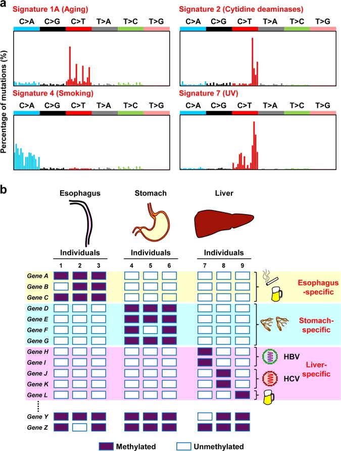
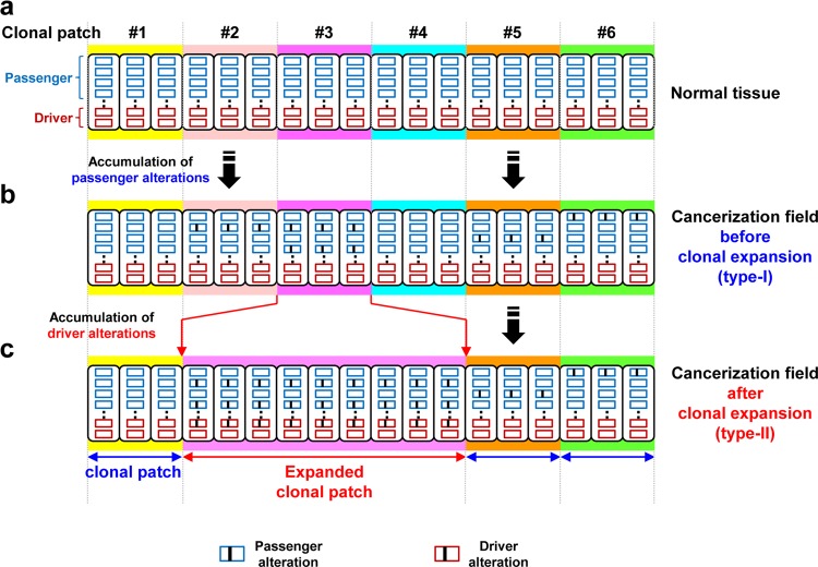
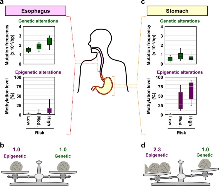
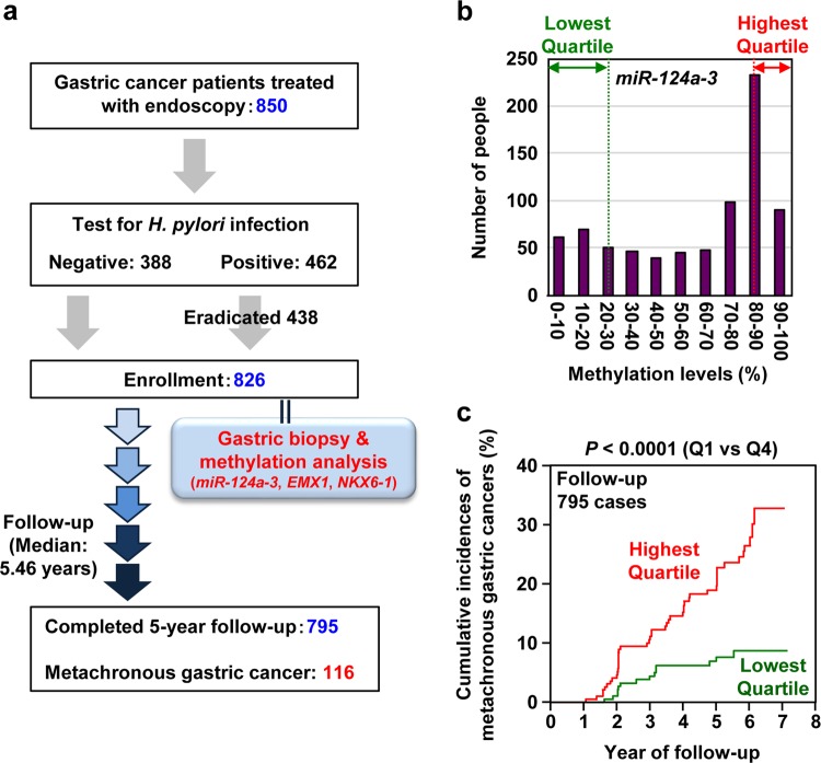
Cancers develop due to the accumulation of genetic and epigenetic alterations. Genetic alterations are induced by aging, mutagenic chemicals, ultraviolet light, and other factors; whereas, epigenetic alterations are mainly by aging and chronic inflammation. The accumulation and patterns of alterations in normal cells reflect our past exposure levels and life history. Most accumulated alterations are considered as passengers, but their accumulation is correlated with cancer drivers. This has been shown for aberrant DNA methylation but has only been speculated for genetic alterations. However, recent technological advancements have enabled measurement of rare point mutations, and studies have shown that their accumulation levels are indeed correlated with cancer risk. When the accumulation levels of aberrant DNA methylation and point mutations are combined, risk prediction becomes even more accurate. When high levels of alterations accumulate, the tissue has a high risk of developing cancer or even multiple cancers and is considered as a "cancerization field", with or without expansion of physiological patches of clonal cells. In this review, we describe the formation of a cancerization field and how we can apply its detection in precision cancer risk diagnosis.


Conflict of interest statement

T.U. is a recipient of funding from Sysmex and Miraca Inc., and has filed a patent for detecting rare point mutations using a small number of sequencing templates.

Figures

Fig. 1
Fig. 1
Reflection of past exposure to carcinogens in genetic and epigenetic alterations. a Reflection in the mutational signature. Exposure to a carcinogen induces a specific mutation signature (adapted from Alexandrov et al.). b Reflection in methylated gene profiles. A different set of genes are aberrantly methylated depending on the tissue types and possibly on the inducers. Unexpressed genes are different among tissues, and specific genes are susceptible to methylation induction in individual tissues, such as Genes A, B, and C in the esophagus, Genes D, E, F, and G in the stomach, and Genes H, I, J, K, and L in the liver. Even among liver-specific susceptible genes, genes are methylated reflecting the cause of inflammation, such as Genes H and I by HBV infection, Genes J and K by HCV infection, and Gene L by alcohol
Fig. 2
Fig. 2
Formation of a field for cancerization. a Normal tissues are assembled from clonal patches of normal cells. b Genetic and epigenetic alterations are potently induced by exposure to specific inducers, and passenger alterations are mainly accumulated in normal tissues without expansion. c Both passenger and driver alterations, which can induce monoclonal cell proliferation, are accumulated in normal tissues with expansion
Fig. 3
Fig. 3
Different impacts of genetic and epigenetic alterations on cancer risk. Both genetic and epigenetic alterations are involved in forming a field for cancerization, but their relative contributions differ depending on the cancer types. a, b The impacts of genetic and epigenetic alterations are similar for ESCCs. Box plots represent median (center line), upper, and lower quartiles (box limits), and 91 and 9 percentiles (whiskers). The original data were obtained from our previous publication. c, d The impact of epigenetic alterations is higher than that of genetic alterations for gastric cancer
Fig. 4
Fig. 4
A multicenter prospective clinical study to predict cancer risk by measuring accumulated alterations. a Study design of the prospective study. Among the 826 patients enrolled, 116 patients developed metachronous gastric cancer after 1 year of enrollment with a median follow-up period of 5.46 years. b Distribution of the DNA methylation level of one of the pre-selected marker genes, miR-124a-3, in non-cancerous gastric tissues. Patients in the highest quartile had DNA methylation levels of 88.1–91.8%, while those in the lowest quartile had those of 8.3–23.0%. c The impact of methylation burden on the risk of metachronous gastric cancer. The highest quartile had a 3-fold higher risk of developing a metachronous gastric cancer than the lowest quartile. Risk prediction among patients who have already been treated for the first cancer is generally very difficult, but was achieved by measuring the methylation burden. This figure was modified from our previous study

References

    1. Rozhok AI, DeGregori J. The evolution of lifespan and age-dependent cancer risk. Trends Cancer. 2016;2:552–560. doi: 10.1016/j.trecan.2016.09.004. - DOI - PMC - PubMed
    1. Tomasetti C, Vogelstein B. Cancer etiology. Variation in cancer risk among tissues can be explained by the number of stem cell divisions. Science. 2015;347:78–81. doi: 10.1126/science.1260825. - DOI - PMC - PubMed
    1. Cogliano VJ, et al. Preventable exposures associated with human cancers. J. Natl. Cancer Inst. 2011;103:1827–1839. doi: 10.1093/jnci/djr483. - DOI - PMC - PubMed
    1. Bykov VJ, Hemminki K. Assay of different photoproducts after UVA, B and C irradiation of DNA and human skin explants. Carcinogenesis. 1996;17:1949–1955. doi: 10.1093/carcin/17.9.1949. - DOI - PubMed
    1. Reardon JT, Bessho T, Kung HC, Bolton PH, Sancar A. In vitro repair of oxidative DNA damage by human nucleotide excision repair system: possible explanation for neurodegeneration in xeroderma pigmentosum patients. Proc. Natl. Acad. Sci. USA. 1997;94:9463–9468. doi: 10.1073/pnas.94.17.9463. - DOI - PMC - PubMed