Associations between vascular risk factors and brain MRI indices in UK Biobank
- PMID: 30854560
- PMCID: PMC6642726
- DOI: 10.1093/eurheartj/ehz100
Associations between vascular risk factors and brain MRI indices in UK Biobank
Abstract
Aims: Several factors are known to increase risk for cerebrovascular disease and dementia, but there is limited evidence on associations between multiple vascular risk factors (VRFs) and detailed aspects of brain macrostructure and microstructure in large community-dwelling populations across middle and older age.
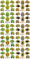
Methods and results: Associations between VRFs (smoking, hypertension, pulse pressure, diabetes, hypercholesterolaemia, body mass index, and waist-hip ratio) and brain structural and diffusion MRI markers were examined in UK Biobank (N = 9722, age range 44-79 years). A larger number of VRFs was associated with greater brain atrophy, lower grey matter volume, and poorer white matter health. Effect sizes were small (brain structural R2 ≤1.8%). Higher aggregate vascular risk was related to multiple regional MRI hallmarks associated with dementia risk: lower frontal and temporal cortical volumes, lower subcortical volumes, higher white matter hyperintensity volumes, and poorer white matter microstructure in association and thalamic pathways. Smoking pack years, hypertension and diabetes showed the most consistent associations across all brain measures. Hypercholesterolaemia was not uniquely associated with any MRI marker.
Conclusion: Higher levels of VRFs were associated with poorer brain health across grey and white matter macrostructure and microstructure. Effects are mainly additive, converging upon frontal and temporal cortex, subcortical structures, and specific classes of white matter fibres. Though effect sizes were small, these results emphasize the vulnerability of brain health to vascular factors even in relatively healthy middle and older age, and the potential to partly ameliorate cognitive decline by addressing these malleable risk factors.
Keywords: Brain; Cortex; Diffusion; MRI; Vascular risk; White matter.
© The Author(s) 2019. Published by Oxford University Press on behalf of the European Society of Cardiology.
Figures


References
-
- Academy of Medical Sciences. Influencing the trajectories of ageing; 2016. https://tinyurl.com/acadmedsci2016 (30 November 2018).
-
- House of Lords. Ageing: Scientific Aspects. London: The Stationery Office; 2005.
-
- Kirkwood T. Foresight Mental Capital and Wellbeing Project 2008: Final Project Report Executive Summary. Mental Capital through Life. London: Government Office for Science; 2008.
-
- Jekel K, Damian N, Wattmo C, Hausner L, Bullock R, Connelly PJ, Dubois PJ, Eriksdotter M, Ewers M, Graessel E, Kramberger MG, Law E, Mecocci P, Molinuevo JL, Nygard L, Olde-Rikkert MG, Orgogozo JM, Pasquier F, Peres K, Salmon E, Sikkes SA, Sobow T, Spiegel R, Tsolaki M, Winblad B, Frolich L.. Mild cognitive impairment and deficits in instrumental activities of daily living: a systematic review. Alzheimers Res Ther 2015;7:17.. - PMC - PubMed
Publication types
MeSH terms
Grants and funding
- MR/J006971/1/MRC_/Medical Research Council/United Kingdom
- MC_UU_12011/2/MRC_/Medical Research Council/United Kingdom
- MR/L023784/2/MRC_/Medical Research Council/United Kingdom
- R01 AG054628/AG/NIA NIH HHS/United States
- BB_/Biotechnology and Biological Sciences Research Council/United Kingdom
- MR/M013111/1/MRC_/Medical Research Council/United Kingdom
- MC_U147585819/MRC_/Medical Research Council/United Kingdom
- G1001245/MRC_/Medical Research Council/United Kingdom
- MR/R024065/1/MRC_/Medical Research Council/United Kingdom
- P2C HD042849/HD/NICHD NIH HHS/United States
- R01 HD083613/HD/NICHD NIH HHS/United States
- G0701120/MRC_/Medical Research Council/United Kingdom
LinkOut - more resources
Full Text Sources
Medical

