. 2019 May;49(5):737-746.
doi: 10.1002/eji.201847949. Epub 2019 Mar 20.

CD1b presents self and Borrelia burgdorferi diacylglycerols to human T cells

Affiliations

CD1b presents self and Borrelia burgdorferi diacylglycerols to human T cells

Peter Reinink et al. Eur J Immunol. 2019 May.

Abstract

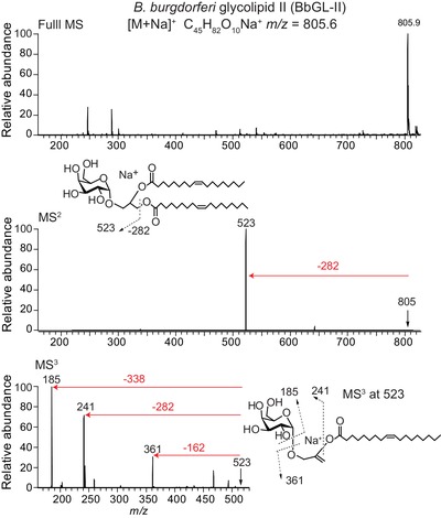
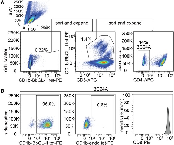
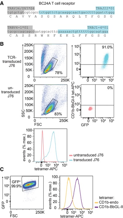
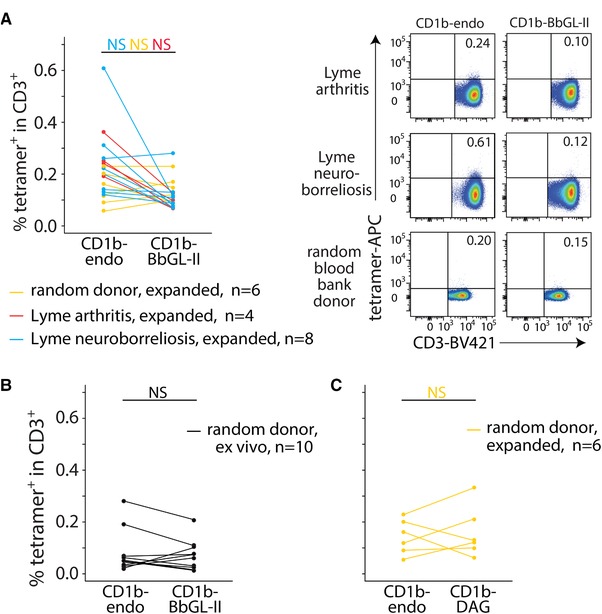
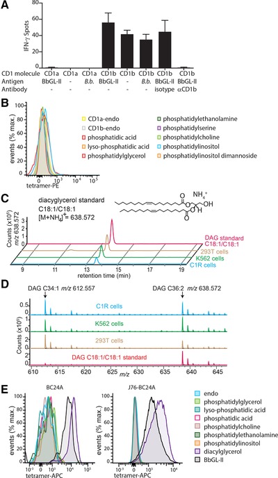
Lyme disease is a common multisystem disease caused by infection with a tick-transmitted spirochete, Borrelia burgdorferi and related Borrelia species. The monoglycosylated diacylglycerol known as B. burgdorferi glycolipid II (BbGL-II) is a major target of antibodies in sera from infected individuals. Here, we show that CD1b presents BbGL-II to human T cells and that the TCR mediates the recognition. However, we did not detect increased frequency of CD1b-BbGL-II binding T cells in the peripheral blood of Lyme disease patients compared to controls. Unexpectedly, mapping the T cell specificity for BbGL-II-like molecules using tetramers and activation assays revealed a concomitant response to CD1b-expressing APCs in absence of BbGL-II. Further, among all major classes of self-lipid tested, BbGL-II responsive TCRs show strong cross-reactivity to diacylglycerol, a self-lipid antigen with structural similarities to BbGL-II. Extending prior work on MHC and CD1b, CD1c, and CD1d proteins, this study provides evidence for cross-reactive CD1b-restricted T cell responses to bacterial and self-antigens, and identifies chemically defined targets for future discovery of self and foreign antigen cross-reactive T cells.

Keywords: CD1b; Lyme disease; T cells; antigen specificity; lipid antigen.


Conflict of interest statement

The authors declare no commercial or financial conflict of interest.

Figures

Figure 1
Figure 1
Mass spectrometry on synthetic BbGL‐II lipid. Positive ion mode ESI–MS of synthetic BbGL‐II gave an expected major ion at m/z 805.9 (top panel), which was subjected to multistage CID‐MS to yield fragment ions as indicated (middle and bottom panels).
Figure 2
Figure 2
Isolation of CD1b‐BbGL‐II binding T cells. (A) PBMC from blood donor BC24 (Table 1) with erythema migrans were sorted based on CD1b‐BbGL‐II tetramer binding. After three rounds of sorting and expansion, the 96% of CD4 T cells that stain with CD1b‐BbGL‐II tetramers were further tested for staining with CD1b‐endo tetramers carrying endogenous (endo) lipids and an antibody against CD8α. Positive CD1b‐BbGL‐II tetramer, negative CD1b‐endo, and negative CD4 staining have been observed in five independent experiments. CD8 staining was performed once.
Figure 3
Figure 3
TCR dependent tetramer binding. (A) The sequence of the CD1b‐BbGL‐II binding TCR was determined by single cell TCR sequencing, where six independently obtained single cells showed the same CDR3 sequence. The nucleotides of the variable region (light grey) N‐region additions (white) and joining segment (dark grey) are color‐coded. (B) J76 cells transduced with the BC24A TCR and untransduced J76 cells were stained with CD1b‐BbGL‐II tetramer. (C) J76 cells transduced with the CD1b‐BbGL‐II specific TCR were stained with CD1b‐BbGL‐II and CD1b‐endo tetramers. Panel (C) was pregated based on forward scatter (FSC) and side scatter (SSC) as shown in (B). Four independent experiments were performed with comparable results. One representative experiment is shown.
Figure 4
Figure 4
Tetramer staining of T cells from Lyme disease patients. T cells that were expanded for 2 weeks (A and C) or PBMC that were used directly ex vivo (B) were stained with an αCD3 antibody and the indicated tetramers (CD1b‐BbGL‐II, CD1b‐endo, or CD1b‐DAG). Facs data of three representative subjects (one Lyme arthritis, one Lyme neuroborreliosis, and one random blood bank donor) are shown in (A). The complete set of flowcytometric data and pregating strategy is shown in Supporting Information Figures 1–5. The percentages CD3+ tetramer+ cells are indicated. NS: not significant (p > 0.05) as calculated by paired Wilcoxon ranked sums tests and Benjamini and Hochberg correction for multiple testing.
Figure 5
Figure 5
CD1b‐BbGL‐II‐binding T cells are autoreactive and recognize diacylglycerol. (A) IFN‐γ production was measured by co‐culture of BC24A T cells and C1R cells transduced to express human CD1a or CD1b. The cells were incubated in the presence of BbGL‐II lipid, sonicated Borrelia burgdorferi (B.b.), or without antigen (–). A blocking antibody or isotype control was added. Data is mean value of triplicates with error bars showing SD. Data shown is representative of three independent experiments. (B) Staining of BC24A with the indicated tetramers. (C) Detection of C36:2 DAG standard or lipid extracts of human cell lines C1R, K562, and 293T cells by high mass resolution mass HPLC time of flight MS. (D) Mass spectrum between 13.5 and 13.7 min shows the presence of C34:1 DAG and C36:2 DAG in cells. (E) Cultured primary T cells or J76 cells transduced with the BC24A TCR were stained with CD1b tetramers loaded with the indicated antigens. Pregating strategy is shown in Figures 2A and 3C, respectively. BC24A staining was performed twice independently, J76‐BC24A staining was performed three times independently. One representative staining is shown.

Similar articles

Cited by

References

    1. Calabi, F. , Jarvis, J. M. , Martin, L. and Milstein, C. , Two classes of CD1 genes. Eur. J. Immunol. 1989. 19: 285–292. - PubMed
    1. Dougan, S. K. , Kaser, A. and Blumberg, R. S. , CD1 expression on antigen‐presenting cells. Curr. Top Microbiol. Immunol. 2007. 314: 113–141. - PubMed
    1. Rossjohn, J. , Gras, S. , Miles, J. J. , Turner, S. J. , Godfrey, D. I. and McCluskey, J. , T cell antigen receptor recognition of antigen‐presenting molecules. Annu. Rev. Immunol. 2015. 33: 169–200. - PubMed
    1. Van Rhijn, I. and Moody, D. B. , Donor unrestricted T cells: A shared human t cell response. J. Immunol. 2015. 195: 1927–1932. - PMC - PubMed
    1. Beckman, E. M. , Porcelli, S. A. , Morita, C. T. , Behar, S. M. , Furlong, S. T. and Brenner, M. B. , Recognition of a lipid antigen by CD1‐restricted alpha beta+ T cells. Nature 1994. 372: 691–694. - PubMed

Publication types

MeSH terms