. 2019 Jun;18(3):e12931.
doi: 10.1111/acel.12931. Epub 2019 Mar 10.

Aged-senescent cells contribute to impaired heart regeneration

Affiliations

Aged-senescent cells contribute to impaired heart regeneration

Fiona C Lewis-McDougall et al. Aging Cell. 2019 Jun.

Abstract

Aging leads to increased cellular senescence and is associated with decreased potency of tissue-specific stem/progenitor cells. Here, we have done an extensive analysis of cardiac progenitor cells (CPCs) isolated from human subjects with cardiovascular disease, aged 32-86 years. In aged subjects (>70 years old), over half of CPCs are senescent (p16INK4A , SA-β-gal, DNA damage γH2AX, telomere length, senescence-associated secretory phenotype [SASP]), unable to replicate, differentiate, regenerate or restore cardiac function following transplantation into the infarcted heart. SASP factors secreted by senescent CPCs renders otherwise healthy CPCs to senescence. Elimination of senescent CPCs using senolytics abrogates the SASP and its debilitative effect in vitro. Global elimination of senescent cells in aged mice (INK-ATTAC or wild-type mice treated with D + Q senolytics) in vivo activates resident CPCs and increased the number of small Ki67-, EdU-positive cardiomyocytes. Therapeutic approaches that eliminate senescent cells may alleviate cardiac deterioration with aging and restore the regenerative capacity of the heart.

Keywords: aging; cardiac regeneration; cardiac repair; myocardial infarction; p16INK4a; progenitor cells; senescence; senescence-associated secretory phenotype; senolytics.


Conflict of interest statement

None declared.

Figures
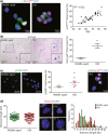
Figure 1
Figure 1
Over half of cardiac progenitor cells (CPCs) in the aged human heart are senescent (a) Representative immunofluorescence images and quantification of c‐kitpos p16INK4Apos CPCs, (n = 35 donors), (b) c‐kitpos SA‐β‐galpos CPCs (*p = 0.0014; n = 2–4), (c) c‐kitpos γ‐H2AXpos p16INK4A‐expressing CPCs (*p = 0.0264; n = 5 donors). (d) Q‐FISH telomere length of single c‐kitpos CPCs (n = 100 cells per group, 20 cells per donor). Representative immunofluorescence images of telomere staining, L5178Y‐S (10 kb) and L5178Y‐R (79 kb) are mouse cell lines with known telomere length. Frequency distribution histogram of CPC telomere length (n = 5 donors/group). Nuclei stained in blue by DAPI. All data are mean ± SEM
Figure 2
Figure 2
Cardiac progenitor cells (CPCs) isolated from aged hearts exhibit diminished proliferation, clonogenicity and cardiomyocyte differentiation potential (a) Quantification of CPC proliferation (*p = 0.0266; n = 3–4 donors), (b) single CPC‐derived clonal efficiency (*p = 0.0008, n = 4–5 donors), (c) CPC spherogenesis number (*p = 0.0051) and (d) size (*p = 0.01), (n = 2–3 donors). (e) Representative immunofluorescence images of undifferentiated and differentiated CPCs from old and middle‐aged donors. Nuclei stained in blue by DAPI. (f) Quantification of Nkx2.5pos (*p = 0.0078; n = 3 donors) and (g) α‐sarcomeric actin expression (*p = 0.0140; n = 3 donors). All data are mean ± SEM
Figure 3
Figure 3
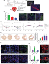
Aged‐senescent CPCs show decreased reparative potential (a) In vivo MI experimental design. (b) Representative confocal images of PKH26pos CPCs engrafted in the myocardium 4 days post‐MI. (c) Quantification and representative confocal images of engraftment of PKH26pos cells 28 days post‐MI (*p = 0.015 vs. c‐kitneg cells, n = 4–5 mice). (d) Echocardiography measurements of LV ejection fraction (EF), fractional shortening (FS), left ventricular end‐diastolic diameter (LVEDD) and left ventricular end‐systolic diameter (LVESD) at baseline (BL) before MI, 7 and 28 days after MI and cell injection (*p < 0.05 vs. Sham, †p < 0.05 vs. Cycling‐CPCs, Δ< 0.05 vs. dox‐induced Sen‐CPCs, n = 5–7 mice). (e) Representative micrographs and quantification of average LV fibrosis (*p < 0.05 vs. Sham; †p = 0.0112 vs. Cycling‐CPCs; n = 5–6 mice). (f) Representative confocal images of PKH26 co‐expression with α‐sarcomeric actinin and vWF (arrowheads) 28 days post‐MI. (g) Quantification and representative confocal images of BrdUpos/α‐sarcomeric actininpos cardiomyocytes and BrdUpos/vWFpos capillaries (*p < 0.05 vs. all; n = 5–7 mice). Nuclei stained in blue by DAPI. All data are mean ± SEM
Figure 4
Figure 4
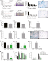
Aged‐senescent CPCs have a SASP (a) Transcript SASP factor expression of dox‐induced Sen‐CPCs relative to Cycling‐CPCs (control). (b) SASP factor protein levels quantified by Luminex of unconditioned media (UM), Cycling‐CPC (CM) and dox‐induced Sen‐CPC (Sen CM) conditioned media (*p < 0.05 vs. UM and CM). Conditioned media applied to cycling‐competent CPCs and the following analyses performed; (c) Quantification and representative staining of CPC proliferation (*p < 0.001; n = 5 replicates), (d) p16INK4A‐pos CPCs (*p < 0.0001; n = 5 replicates), (e) SA‐β‐galpos CPCs (*p < 0.0001; n = 5 replicates), (f) γH2AXpos CPCs (*p < 0.001; n = 5 replicates). All data are mean ± SEM
Figure 5
Figure 5
Senolytic clearance abrogates the SASP (a) Viability (Crystal violet) and SA‐β‐gal quantification of dox‐induced Sen‐CPCs and Cycling‐CPCs exposed to various concentrations of D + Q for 3 days. (b–f) Quantification of 7 days of co‐culture of Cycling‐CPCs with dox‐induced Sen‐CPCs for (b) viability (*p = 0.0068); (c) proliferation (*p = 0.0056); (d) p16INK4A (*p = 0.0025); (e) SA‐β‐gal (*p = 0.0025); (f) γH2AX (*p = 0.0002). CTRL is Cycling‐ CPCs alone. (n = 5 replicates). (g) Representative SA‐β‐gal staining after clearance of dox‐induced Sen‐CPCs from co‐culture by D + Q treatment. (h–k) Quantification of 17 days of co‐culture of Cycling‐CPCs with dox‐induced Sen‐CPCs or co‐culture with D + Q treatment for (h) viability (*p < 0.0001 vs. CTRL 17 days; †p = 0.0001 vs. co‐culture 17 days); (i) proliferation (*p < 0.0001 vs. CTRL 17 days; †p < 0.0001 vs. co‐culture 17 days); (j) p16INK4A (*p < 0.0001 vs. CTRL 17 days; †p < 0.0001 vs. co‐culture 17 days) and (k) SA‐β‐gal (*p < 0.0001 vs. CTRL 17 days; †p < 0.0001 vs. co‐culture 17 days). CTRL is Cycling‐CPCs alone. (n = 5 replicates). (l) SASP factor protein levels quantified by Luminex from each treatment condition (*p < 0.0001 vs. CTRL; †p < 0.01 vs. co‐culture 7 days and 17 days). (n = 2 replicates). All data are mean ± SEM
Figure 6
Figure 6
Clearance of senescent cells stimulates new cardiomyocyte formation in the aged heart (a) In vivo senescent cell clearance experimental design. (b) Total cardiac p16Ink4a gene expression following clearance (*p < 0.01 vs. Vehicle; n = 5 mice). (c) Quantification of CPCs following clearance, (*p < 0.0001 vs. Vehicle; †p = 0.0453 vs. AP; n = 10–11 mice). (d) Frequency distribution histogram of cardiomyocyte diameter, (n = 6 mice). (e) A Ki67pos/α‐sarcomeric actinpos cardiomyocyte (arrowhead) in the LV of a 32 month D + Q‐treated mouse. (f) Quantification of Ki67pos cardiomyocytes, (*p < 0.0001 vs. Vehicle; n = 10 mice). (g) An EdUpos/α‐sarcomeric actinpos cardiomyocyte in the LV of a 22 month INK‐ATTAC AP‐treated mouse. Nuclei are stained by DAPI in blue. (h) Quantification of EdUpos cardiomyocytes (*p < 0.0001 vs. Old + Vehicle; †p < 0.0001 vs. Young + AP; n = 5 mice). (i) Quantification of LV fibrosis (*p < 0.05 vs. Vehicle; n = 3 mice). All data are mean ± SD

References

    1. Baker, D. J. , Childs, B. G. , Durik, M. , Wijers, M. E. , Sieben, C. J. , Zhong, J. , … van Deursen, J. M. (2016). Naturally occurring p16Ink4a‐positive cells shorten healthy lifespan. Nature, 530(7589), 184–189. 10.1038/nature16932 - DOI - PMC - PubMed
    1. Baker, D. J. , Wijshake, T. , Tchkonia, T. , LeBrasseur, N. K. , Childs, B. G. , van de Sluis, B. , … van Deursen, J. M. (2011). Clearance of p16Ink4a‐positive senescent cells delays ageing‐associated disorders. Nature, 479, 232 10.1038/nature10600 - DOI - PMC - PubMed
    1. Bergmann, O. , Bhardwaj, R. D. , Bernard, S. , Zdunek, S. , Barnabe‐Heider, F. , Walsh, S. , … Frisen, J. (2009). Evidence for cardiomyocyte renewal in humans. Science, 324(5923), 98–102. 10.1126/science.1164680 - DOI - PMC - PubMed
    1. Broughton, K. M. , Wang, B. J. , Firouzi, F. , Khalafalla, F. , Dimmeler, S. , Fernandez‐Aviles, F. , & Sussman, M. A. (2018). Mechanisms of cardiac repair and regeneration. Circulation Research, 122(8), 1151–1163. 10.1161/circresaha.117.312586 - DOI - PMC - PubMed
    1. Canela, A. , Vera, E. , Klatt, P. , & Blasco, M. A. (2007). High‐throughput telomere length quantification by FISH and its application to human population studies. Proceedings of the National Academy of Sciences of the United States of America, 104(13), 5300–5305. 10.1073/pnas.0609367104 - DOI - PMC - PubMed

Publication types