

Hypertrophic Obstructive Cardiomyopathy
- PMID: 30855006
- PMCID: PMC6415619
- DOI: 10.3238/arztebl.2019.0047
Hypertrophic Obstructive Cardiomyopathy
Abstract
Background: Hypertrophic cardiomyopathy (HCM) is caused by mutations in a number of genes. Its prevalence is 0.2% to 0.6%.
Methods: This review is based on publications retrieved by a selective literature search and on the authors' clinical experi- ence.
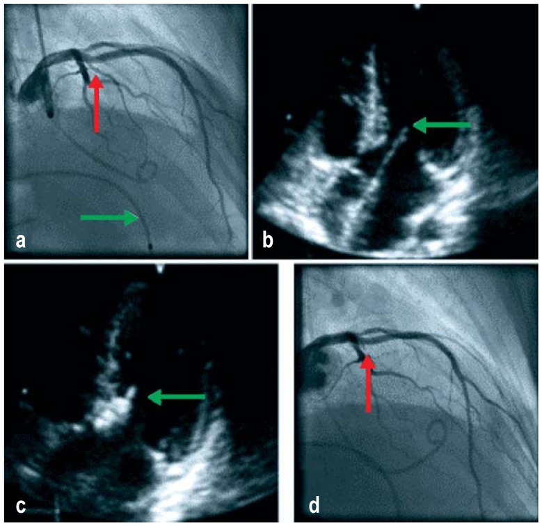
Results: 70% of patients with HCM suffer from the obstructive type of the condition, clinically characterized by highly dynamic and variable manifestations in the form of dyspnea, angina pectoris, and stress-dependent presyncope and syn- cope. Younger patients are at particular risk of sudden cardiac death; thus, all patients need not only symptomatic treatment, but also risk assessment, which can be difficult in individual cases. Left ventricular obstruction, which usually causes symptoms, is treated medically at first, with either a beta- blocker or verapamil. If medical treatment fails, two invasive treatments are available, surgical myectomy and percu- taneous septum ablation. Both of these require a high level of expertise. If performed successfully, they lead to sustained gradient reduction and clinical improvement. Septum ablation is associated with low perioperative and peri-interventional mortality but necessitates permanent pacemaker implantation in 10-20% of patients.
Conclusion: In the absence of evidence from randomized comparison trials, a suitable method of reducing the gradient should be determined by an HCM team in conjunction with each individual patient. Important criteria for decision-making include the anatomical findings and any accompanying illnesses.
Figures


References
-
- Elliott PM, Anastasakis A, et al. Authors/Task Force members. 2014 ESC guidelines on diagnosis and management of hypertrophic cardiomyopathy: the Task Force for the Diagnosis and Management of Hypertrophic Cardiomyopathy of the European Society of Cardiology (ESC) Eur Heart J. 2014;35:2733–2779. - PubMed
-
- Nishimura RA, Holmes DR. Clinical practice Hypertrophic obstructive cardiomyopathy. N Engl J Med. 2004;350:1320–1327. - PubMed
-
- Maron BJ, Gardin JM, Flack JM, Gidding SS, Kurosaki TT, Bild DE. Prevalence of hypertrophic cardiomyopathy in a general population of young adults Echocardiographic analysis of 4111 subjects in the CARDIA study. Coronary artery risk development in (young) adults. Circulation. 1995;92:785–789. - PubMed
-
- Semsarian C, Ingles J, Maron MS, Maron BJ. New perspectives on the prevalence of hypertrophic cardiomyopathy. J Am Coll Cardiol. 2015;65:1249–1254. - PubMed
-
- Koljaja-Batzner A, Pfeiffer B, Seggewiss H. Die hypertrophe Kardiomyopathie - häufig und nicht erkannt. Internistische Praxis. 2018;59:187–201.
Publication types
MeSH terms
LinkOut - more resources
Full Text Sources
Miscellaneous