. 2019 Jun 1;76(6):690-700.
doi: 10.1001/jamaneurol.2019.0204.

Association of Piriform Cortex Resection With Surgical Outcomes in Patients With Temporal Lobe Epilepsy

Affiliations

Association of Piriform Cortex Resection With Surgical Outcomes in Patients With Temporal Lobe Epilepsy

Marian Galovic et al. JAMA Neurol. .

Abstract

Importance: A functional area associated with the piriform cortex, termed area tempestas, has been implicated in animal studies as having a crucial role in modulating seizures, but similar evidence is limited in humans.

Objective: To assess whether removal of the piriform cortex is associated with postoperative seizure freedom in patients with temporal lobe epilepsy (TLE) as a proof-of-concept for the relevance of this area in human TLE.

Design, setting, and participants: This cohort study used voxel-based morphometry and volumetry to assess differences in structural magnetic resonance imaging (MRI) scans in consecutive patients with TLE who underwent epilepsy surgery in a single center from January 1, 2005, through December 31, 2013. Participants underwent presurgical and postsurgical structural MRI and had at least 2 years of postoperative follow-up (median, 5 years; range, 2-11 years). Patients with MRI of insufficient quality were excluded. Findings were validated in 2 independent cohorts from tertiary epilepsy surgery centers. Study follow-up was completed on September 23, 2016, and data were analyzed from September 24, 2016, through April 24, 2018.

Exposures: Standard anterior temporal lobe resection.

Main outcomes and measures: Long-term postoperative seizure freedom.

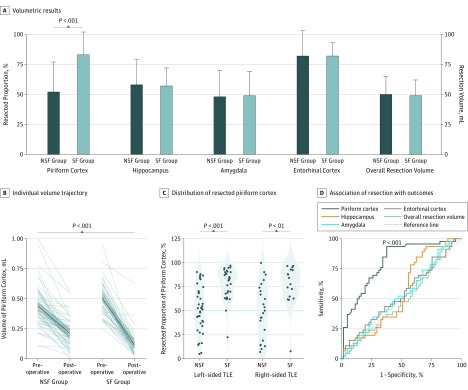
Results: In total, 107 patients with unilateral TLE (left-sided in 68; 63.6% women; median age, 37 years [interquartile range {IQR}, 30-45 years]) were included in the derivation cohort. Reduced postsurgical gray matter volumes were found in the ipsilateral piriform cortex in the postoperative seizure-free group (n = 46) compared with the non-seizure-free group (n = 61). A larger proportion of the piriform cortex was resected in the seizure-free compared with the non-seizure-free groups (median, 83% [IQR, 64%-91%] vs 52% [IQR, 32%-70%]; P < .001). The results were seen in left- and right-sided TLE and after adjusting for clinical variables, presurgical gray matter alterations, presurgical hippocampal volumes, and the proportion of white matter tract disconnection. Findings were externally validated in 2 independent cohorts (31 patients; left-sided TLE in 14; 54.8% women; median age, 41 years [IQR, 31-46 years]). The resected proportion of the piriform cortex was individually associated with seizure outcome after surgery (derivation cohort area under the curve, 0.80 [P < .001]; external validation cohorts area under the curve, 0.89 [P < .001]). Removal of at least half of the piriform cortex increased the odds of becoming seizure free by a factor of 16 (95% CI, 5-47; P < .001). Other mesiotemporal structures (ie, hippocampus, amygdala, and entorhinal cortex) and the overall resection volume were not associated with outcomes.

Conclusions and relevance: These results support the importance of resecting the piriform cortex in neurosurgical treatment of TLE and suggest that this area has a key role in seizure generation.


Conflict of interest statement

Conflict of Interest Disclosures: Dr Galovic reported grant MR/L013215/1 from the Medical Research Council (MRC) outside the submitted work. Dr Baxendale reported donating honoraria from Eisai to the International League Against Epilepsy. Dr Nachev reported grants from NIHR and Wellcome Trust during the conduct of the study. Dr Vos reported receiving grants from UCL Hospital (UCLH)/UCL, National Institute of Health Research (NIHR) Biomedical Research Center (BRC), during the conduct of the study. Dr Winston reported grants from the MRC, NIHR, and Sobell Foundation during the conduct of the study. Dr Wu reported consulting with Medtronic, Inc, Neuropace, Inc, Nevro Corp, and Micro Systems Engineering. Dr Davis reported grants from Eisai outside the submitted work and award K23-NS0929373: K award from the National Institute of Neurological Disorders and Stroke (NINDS) (principal investigator). Dr Litt reported nonfinancial support from Blackfynn, Inc; grants from the NIH (No. R01 NS099348-01) and The Mirowski Family Foundation, as well as personal fees from EpilepsyCo outside the submitted work; and involvement in innovation and licensing intellectual property to Blackfynn Inc, NeuroPace Inc, EpilepsyCo and MC10 Inc, that look to develop new diagnostic and therapeutic technologies for epilepsy and do not pertain directly to the submitted work. Dr Moshé reported grants from the NINDS and grants from the Department of Defense during the conduct of the study and personal fees from UCB, Mallinkrodt, and Eisai outside the submitted work. Dr Sander reported grants and personal fees from UCB and Eisai, personal fees from Janssen, and grants from GW Pharmaceuticals outside the submitted work. Dr Löscher reported grants from the German Research Foundation (Lo274/15-1 and 16-1) and from the European Union’s Seventh Framework Programme under grant agreement 602102 (EPITARGET) during the conduct of the study. Dr Duncan reported grants from NIHR, MRC, and Wellcome Trust during the conduct of the study. Dr Koepp reported grants from MRC and Wellcome Trust during the conduct of the study. No other disclosures were reported.

Figures

Figure 1.
Figure 1.. Localization of the Piriform Cortex and Voxel-Based Morphometry (VBM) Results
The position of the piriform cortex relative to other mesiotemporal structures is illustrated using 3-dimensional (3D) reconstructions (A) and magnetic resonance imaging (MRI) sections (B). Numbers are Montreal Neurological Institute (MNI) section coordinates. The remnant of the piriform cortex is outlined on postsurgical MRI scans of 2 cases (C). The left image is from a right-handed woman in her 30s with incomplete resection of the piriform cortex who continued having seizures after epilepsy surgery. The right image is from a right-handed man in his 40s with more extensive resection of the piriform cortex who became seizure free. Both cases had right-sided temporal lobe epilepsy (TLE) with neuropathologically confirmed hippocampal sclerosis. The statistical parametric maps for the interaction between seizure-free (SF) vs non–seizure-free (NSF) groups and preoperative vs postoperative scans are displayed in the lower half of the Figure (D). This comparison allows determination of gray matter decreases due to epilepsy surgery (ie, comparing postoperative with preoperative images) that were present in SF but not in NSF participants. The statistical mask of significant gray matter volume decreases (red; threshold, uncorrected P < .001) for left- (n = 68) and right-sided (n = 39) TLE is superimposed on coronal (top), axial (middle), and sagittal (bottom) views of a standard MRI template (numbers below are MNI section coordinates). The probability of a voxel being surgically resected is overlaid in blue (range, 10%-100%). Also displayed are zoomed sections in rectangles and 3D reconstructions of neocortical resection margins.
Figure 2.
Figure 2.. Volumetric Results in the Derivation Cohort of Patients Undergoing Surgery for Temporal Lobe Epilepsy (TLE)
Volumetric results (A) are displayed as median (SD) (error bars) of the resected proportion of the piriform cortex, hippocampus, amygdala, and entorhinal cortex and the overall resection volume in the overall cohort (n = 107). Comparing postoperatively seizure-free (SF [n = 46]) with non–seizure-free (NSF [n = 61]) groups, difference in piriform cortex volumes was significant, whereas no differences were observed in all other regions. Gray lines show the individual trajectories of presurgical vs postsurgical volumes of the piriform cortex in the SF vs NSF groups (B). The mean trajectory is illustrated by a dark blue line; SD, by the light blue area. The distribution of the resected proportion of the piriform cortex in the NSF vs SF groups is shown (C); the 50% resection cutoff is displayed as a blue horizontal line. The association of resection area with outcomes (D) used receiver operating characteristics curves to describe individual discrimination. The piriform cortex was the only area to significantly discriminate between SF and NSF groups in the derivation cohort. All other areas remained close to the 45° reference line.
Figure 3.
Figure 3.. Volumetric Results in the Validation Cohort
Gray lines show the individual trajectories of presurgical vs postsurgical volumes of the piriform cortex in the seizure-free (SF) vs non–seizure-free (NSF) groups (A). The mean trajectory is illustrated by a dark blue line; SD, by the light blue area. The distribution of the resected proportion of the piriform cortex is compared in the NSF and SF groups (B). The 50% resection cutoff is displayed as a blue horizontal line. The association of piriform cortex resection with outcome (C) used receiver operating characteristics curves to describe individual-subject discrimination. TJU indicates Thomas Jefferson University; UP, University of Pennsylvania.

References

    1. de Tisi J, Bell GS, Peacock JL, et al. . The long-term outcome of adult epilepsy surgery, patterns of seizure remission, and relapse: a cohort study. Lancet. 2011;378(9800):1388-1395. doi:10.1016/S0140-6736(11)60890-8 - DOI - PubMed
    1. Najm I, Jehi L, Palmini A, Gonzalez-Martinez J, Paglioli E, Bingaman W. Temporal patterns and mechanisms of epilepsy surgery failure. Epilepsia. 2013;54(5):772-782. doi:10.1111/epi.12152 - DOI - PubMed
    1. Piredda S, Gale K. A crucial epileptogenic site in the deep prepiriform cortex. Nature. 1985;317(6038):623-625. doi:10.1038/317623a0 - DOI - PubMed
    1. Gale K. Progression and generalization of seizure discharge: anatomical and neurochemical substrates. Epilepsia. 1988;29(suppl 2):S15-S34. doi:10.1111/j.1528-1157.1988.tb05795.x - DOI - PubMed
    1. McIntyre DC, Kelly ME. The parahippocampal cortices and kindling. Ann N Y Acad Sci. 2000;911:343-354. doi:10.1111/j.1749-6632.2000.tb06736.x - DOI - PubMed

Publication types

MeSH terms