. 2019;8(2):129-143.
doi: 10.3233/JHD-180312.

Cysteamine Protects Neurons from Mutant Huntingtin Toxicity

Affiliations

Cysteamine Protects Neurons from Mutant Huntingtin Toxicity

Nicolas Arbez et al. J Huntingtons Dis. 2019.

Erratum in

Abstract

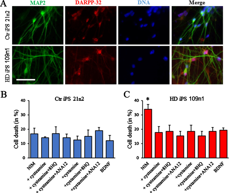
Background: The potential benefit of cysteamine for Huntington's disease has been demonstrated in HD animal models. Cysteamine and its derivate cystamine were shown to reduce neuropathology and prolong lifespan. Human studies have demonstrated safety, and suggestive results regarding efficacy. Despite all the studies available in vivo, there are only few in vitro studies, and the mechanism of action of cysteamine remains unclear.

Objective: The objective of this study is to assess the capacity of cysteamine for neuroprotection against mutant Huntingtin in vitro using cellular models of HD, and to provide initial data regarding mechanism of action.

Methods: We tested the neuroprotective properties of cysteamine in vitro in our primary neuron and iPSC models of HD.

Results: Cysteamine showed a strong neuroprotective effect (EC50 = 7.1 nM) against mutant Htt-(aa-1-586 82Q) toxicity, in a nuclear condensation cell toxicity assay. Cysteamine also rescued mitochondrial changes induced by mutant Htt. Modulation of the levels of cysteine or glutathione failed to protect neurons, suggesting that cysteamine neuroprotection is not mediated through cysteine metabolism. Taurine and Hypotaurine, which are metabolites of cysteamine can protect neurons against Htt toxicity, but the inhibition of the enzyme converting cysteamine to hypotaurine does not block either protective activity, suggesting independent protective pathways. Cysteamine has been suggested to activate BDNF secretion; however, cysteamine protection was not blocked by BDNF pathway antagonists.

Conclusions: Cysteamine was strongly neuroprotective with relatively high potency. We demonstrated that the main neuroprotective pathways that have been proposed to be the mechanism of protection by cysteamine can all be blocked and still not prevent the neuroprotective effect. The results suggest the involvement of other yet-to-be-determined neuroprotective pathways.

Keywords: Huntington’s disease; cysteamine; huntingtin toxicity; neurodegeneration; neuroprotection; patient-derived induced pluripotent stem cells; primary neurons.


Conflict of interest statement

CONFLICT OF INTEREST

The authors declare that they have no conflict of interest.

Figures

Fig. 1.
Fig. 1.
Cysteamine protects primary cortical neurons from mHtt toxicity. Primary cortical neurons were transfected at 7 DIV with either Htt N586 82Q or Htt N586 22Q and treated with different doses of cysteamine. After 48 h, neurons were fixed and stained with DAPI and MAP2 was immunodetected. A) Representative pictures of transfected neurons. Scale bar: 25 μm. B) mHtt toxicity measured by nuclear condensation assay was reduced by cysteamine, Results presented as means ± S.E. of the percentage of dead cells. *p < 0.05 vs Htt N586 82Q by ANOVA with Bonferroni post-hoc test. (n = 4 independent neuronal preparations).
Fig. 2.
Fig. 2.
Cysteamine protects from morphological changes induced by mHtt. Primary cortical neurons were transfected at 7 DIV with either Htt N586 82Q, Htt N586 22Q or GFP alone and treated with 1 μM cysteamine. A) Representative images of GFP transfected neurons at 0, 5, and 10 h of live cell imaging. Scale bar: 50 μm. B) Quantification of survival over time by time lapse imaging morphological analysis. Transfected cells were identified live on the microscope and imaged every 15 min for 12 h. Cells are evaluated as 100 if alive and 0 dead. Results are presented as means ± S.E. of cell survival over time (n = 100 individual neurons from N = 5 independent experiments).
Fig. 3.
Fig. 3.
Cysteamine protects primary striatum neurons from mitochondria impairments induced by mHtt. A, B) Measurement of mitochondrial potential using the potential sensitive mitochondrial dye TMRM staining. 24 h after transfections, cells were loaded with 100 nM TMRM for 45 min before individual transfected cells were imaged. A) Examples of cells stained with TMRM. Scale bar: 10 μm. B) Intensity quantification was done using Volocity and the data was normalized to the GFP only transfected cells. Results presented as means ± S.E. of normalized potential. *p < 0.05 vs Htt N586 22Q; #p < 0.05 vs Htt N586 82Q by ANOVA with Bonferroni post-hoc test. (n = 6 independent neuronal preparations). C, D) Measurement of mitochondrial size using Mitotracker. 24 h after transfections, cells were loaded for 45 min with 500 nM Mitotracker for 45 min before fixation and individual transfected cells were imaged. A) Examples of cells stained with Mitotracker. Scale bar: 10 μm. B) Size quantification was done using Volocity and the data was normalized to the GFP only transfected cells. Results presented as means ± S.E. of normalized size. *p < 0.05 vs Htt N586 22Q; #p < 0.05 vs Htt N586 82Q by ANOVA with Bonferroni post-hoc test. (n = 6 independent neuronal preparations).
Fig. 4.
Fig. 4.
Cysteamine protects primary striatum neurons from mHtt toxicity. Primary striatal neurons were transfected at 7 DIV with either Htt N586 82Q or Htt N586 22Q and treated with cysteamine. Htt toxicity was reduced by cysteamine treatment as measured by (A) nuclear condensation, (B) measurement of mitochondrial potential using TMRM staining and (C) measurement of mitochondrial size using Mitotracker. *p < 0.05 vs N86–22Q, #p < 0.05 vs N586–82Q by ANOVA with Bonferroni post-hoc test. (n = 4 independent neuronal preparations).
Fig. 5.
Fig. 5.
Cysteamine metabolites protection from mHtt toxicity. Primary cortical neurons were transfected at 7 DIV with either Htt N586 82Q or Htt N586 22Q and treated with cell metabolites. A) From the cysteine pathway. Either 1 mM L-cysteine, D-cysteine, NAc-L-cysteine or GSH for 48 h. B) From the Taurine pathway. Either 1 μM cysteamine, hypotaurine or taurine in presence or absence of 5 mM 8HQ for 48 h. Cell death was measured by nuclear condensation assay. *p < 0.05 vs N586–22Q by ANOVA with Bonferroni post-hoc test. (n = 4 independent neuronal preparations).
Fig. 6.
Fig. 6.
Cysteamine neuroprotection does not require BDNF pathway activation. Primary cortical neurons were transfected at 7 DIV with either Htt N586 82Q or Htt N586 22Q and treated with either 1 μM cysteamine or 20 ng/mL BDNF. Cell death was measured by nuclear condensation assay after 48 h with cotreatment with 10 μM ANA12 (A) or 1 μM K252a (B). *p < 0.05 vs N586–22Q by ANOVA with Bonferroni post-hoc test. (n = 6 independent neuronal preparations).
Fig. 7.
Fig. 7.
Cystamine protects primary cortical neurons from mHtt toxicity. Primary cortical neurons were transfected at 7 DIV with either Htt N586 82Q or Htt N586 22Q with additional treatments. Cell death was measured at 48 h by nuclear condensation assay. A) Dose response of cysteamine neuroprotection. B) Cystamine protection in presence or absence of the cysteamine dioxygenase 8HQ (5 mM). C) Cystamine protection in presence or absence of the TrkB antagonist ANA12. *p < 0.05 vs Htt N586 82Q by ANOVA with Bonferroni post-hoc test. (n = 4 independent neuronal preparations).
Fig. 8.
Fig. 8.
Cysteamine neuroprotection in HD iPS cells. 21n2 control cells and 109n1 HD iPS cells were differentiated into medium spiny neurons according to our protocol. A) Examples of immunostaining of control and HD cells showing the expression of MAP2 and DARP32. To measure toxicity, differentiated 21n2 control cells (B) and 109n1 HD iPS cells (C) were cultivated in NIM medium for 48 h. Cysteamine and other compounds were added to NIM for all time of BDNF withdrawal. 20 ng/ml BDNF were used as a positive control. Cell death was quantified by nuclear condensation assay. *p < 0.05 vs BDNF by ANOVA with Bonferroni post-hoc test. (n = 4 independent neuronal preparations).

References

    1. McColgan P, Tabrizi SJ. Huntington’s disease: A clinical review. Eur J Neurol. 2018;25(1):24–34. - PubMed
    1. Besouw M, Masereeuw R, van den Heuvel L, Levtchenko E. Cysteamine: An old drug with new potential. Drug Discov Today. 2013;18(15–16):785–92. - PubMed
    1. Dedeoglu A, Kubilus JK, Jeitner TM, Matson SA, Bogdanov M, Kowall NW, et al. Therapeutic effects of cystamine in a murine model of Huntington’s disease. J Neurosci. 2002;22(20):8942–50. - PMC - PubMed
    1. Karpuj MV, Becher MW, Steinman L. Evidence for a role for transglutaminase in Huntington’s disease and the potential therapeutic implications. Neurochem Int. 2002;40(1):31–6. - PubMed
    1. Van Raamsdonk JM, Pearson J, Bailey CD, Rogers DA, Johnson GV, Hayden MR, et al. Cystamine treatment is neuroprotective in the YAC128 mouse model of Huntington disease. J Neurochem. 2005;95(1):210–20. - PubMed

Publication types

LinkOut - more resources