. 2019 Mar 8;9(1):39.
doi: 10.3390/bios9010039.

An Exfoliated Graphite-Based Electrochemical Immunosensor on a Dendrimer/Carbon Nanodot Platform for the Detection of Carcinoembryonic Antigen Cancer Biomarker

Affiliations

An Exfoliated Graphite-Based Electrochemical Immunosensor on a Dendrimer/Carbon Nanodot Platform for the Detection of Carcinoembryonic Antigen Cancer Biomarker

Azeez O Idris et al. Biosensors (Basel). .

Abstract

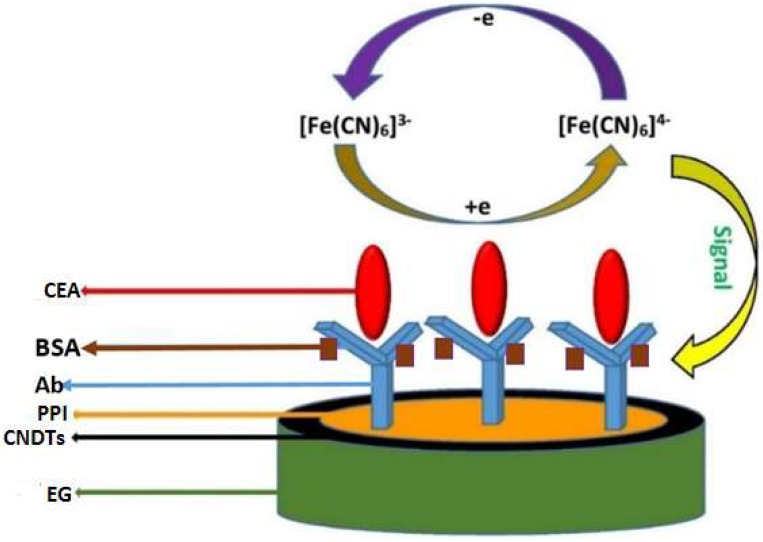
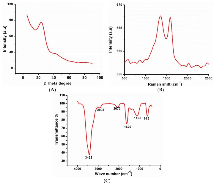
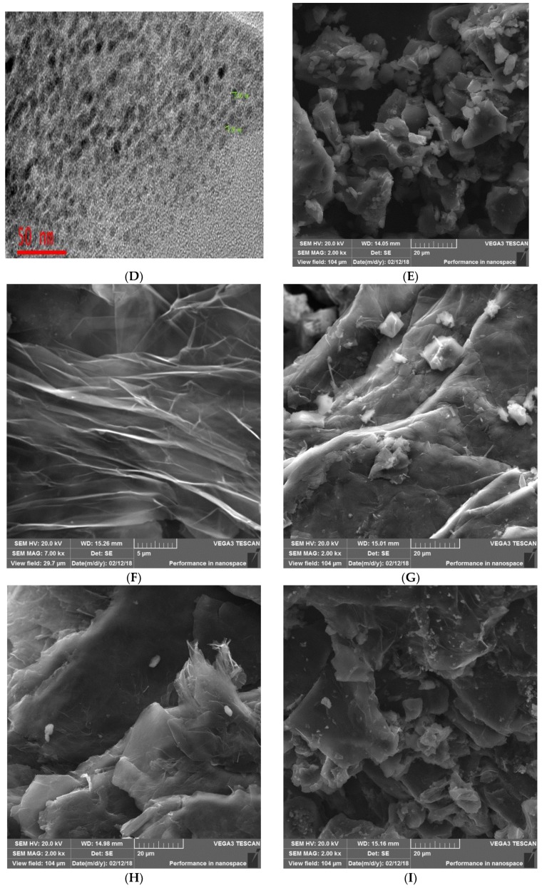
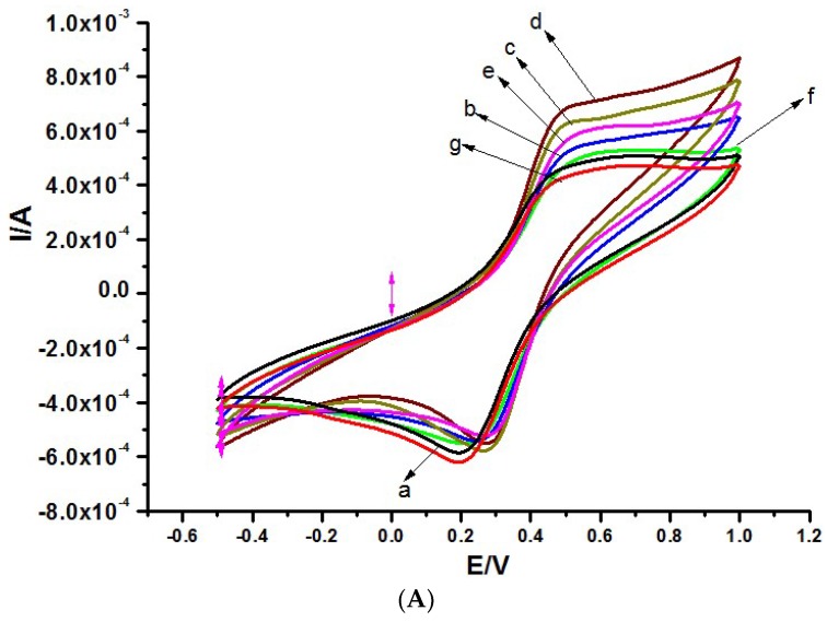
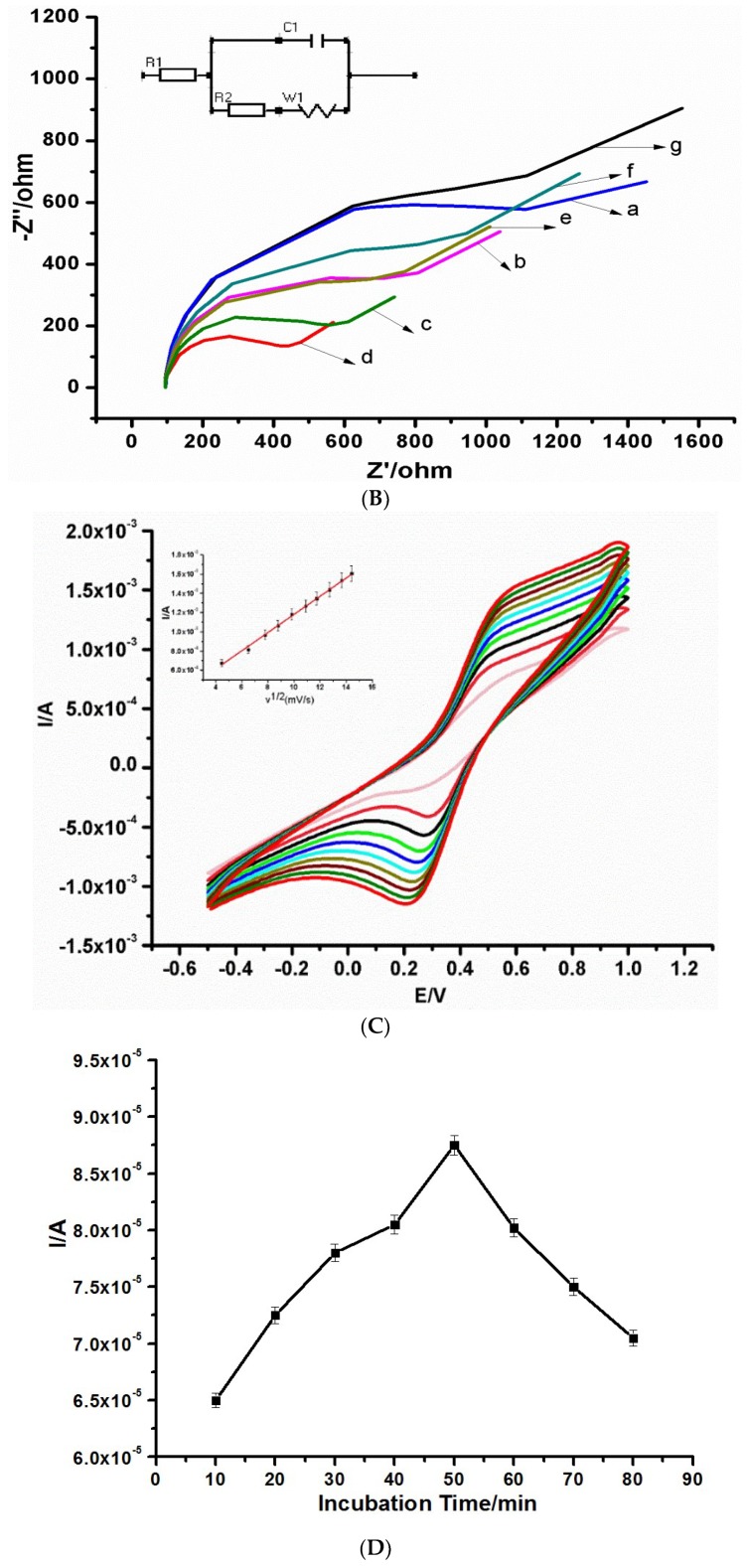
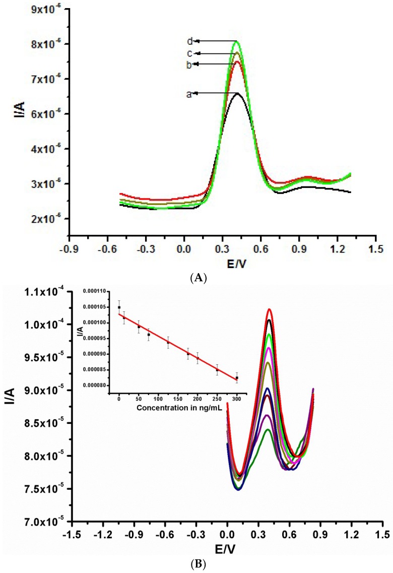
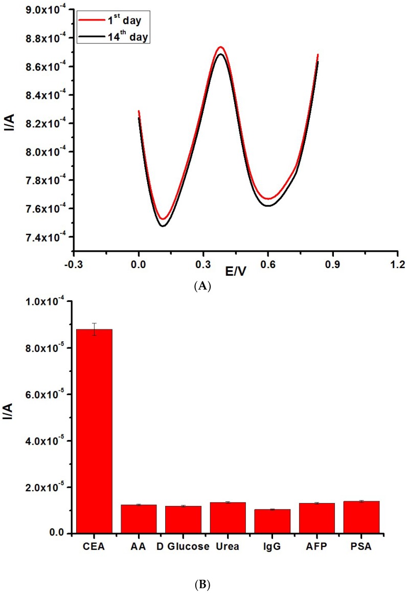
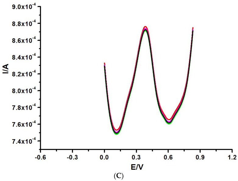
An electrochemical immunosensor for the quantification of carcinoembryonic antigen (CEA) using a nanocomposite of polypropylene imine dendrimer (PPI) and carbon nanodots (CNDTs) on an exfoliated graphite electrode (EG) is reported. The carbon nanodots were prepared by pyrolysis of oats. The nanocomposites (PPI and CNDTs) were characterized using X-ray powder diffraction (XRD), Raman spectroscopy, Fourier transform infrared spectroscopy (FTIR), high-resolution transmission electron microscopy (HRTEM) and scanning electron microscopy (SEM). The proposed immunosensor was prepared on an exfoliated graphite electrode sequentially by drop coating CNDTs, the electrodeposition of G2-PPI (generation 2 poly (propylene imine) dendrimer), the immobilization of anti-CEA on the modified electrode for 80 min at 35 °C, and dropping of bovine serum albumin (BSA) to minimize non-specific binding sites. Cyclic voltammetry was used to characterize each stage of the fabrication of the immunosensor. The proposed immunosensor detected CEA within a concentration range of 0.005 to 300 ng/mL with a detection limit of 0.00145 ng/mL by using differential pulse voltammetry (DPV). The immunosensor displayed good stability and was also selective in the presence of some interference species such as ascorbic acid, glucose, alpha-fetoprotein, prostate-specific antigen and human immunoglobulin. Furthermore, the fabricated immunosensor was applied in the quantification of CEA in a human serum sample, indicating its potential for real sample analysis.

Keywords: cancer; carcinoembryonic antigen; exfoliated graphite electrode; immunosensor; polypropylene imine.


Conflict of interest statement

The authors declare no conflict of interest.

Figures

Scheme 1
Scheme 1
Processes involved in the preparation of the immunosensor.
Figure 1
Figure 1
(A) XRD spectrum of carbon nanodots (CNDTs). (B) Raman spectrum of CNDTs. (C) Fourier transform infrared spectroscopy (FTIR) of CNDTs. (D) TEM image of CNDTs. (E) SEM image of CNDTs. (F) SEM image of the exfoliated graphite electrode. (G) SEM image of CNDTs deposited on exfoliated graphite electrode. (H) SEM image of G2-PPI electrodeposited on the exfoliated graphite electrode. (I) SEM image of CNDTs@G2-PPI on the exfoliated graphite electrode.
Figure 1
Figure 1
(A) XRD spectrum of carbon nanodots (CNDTs). (B) Raman spectrum of CNDTs. (C) Fourier transform infrared spectroscopy (FTIR) of CNDTs. (D) TEM image of CNDTs. (E) SEM image of CNDTs. (F) SEM image of the exfoliated graphite electrode. (G) SEM image of CNDTs deposited on exfoliated graphite electrode. (H) SEM image of G2-PPI electrodeposited on the exfoliated graphite electrode. (I) SEM image of CNDTs@G2-PPI on the exfoliated graphite electrode.
Figure 2
Figure 2
(A) CV and (B) EIS of (a) Bare exfoliated graphite electrode (EG), (b) EG/PPI, (c) EG/CNDTs, (d) EG/CNDTs@PPI, (e) EG/CNDTs@PPI/Anti-CEA, (f) EG/CNDTs@PPI/Anti-CEA/BSA, and (g) EG/CNDTs@PPI/Anti-CEA/BSA/CEA. (C) CV of different scan rates of immunosensor (EG/CNDTs@PPI/Anti-CEA/BSA) in solution containing 1 mM [Fe(CN)6]3−/4− prepared in 0.1 M KCl. The inset is a plot of current versus the square root of scan rates. (D) Optimization of CEA detection incubation time. (E) Optimization of CEA detection incubation temperature in solution containing 1 mM [Fe(CN)6]3−/4− prepared in 0.1 M KCl.
Figure 2
Figure 2
(A) CV and (B) EIS of (a) Bare exfoliated graphite electrode (EG), (b) EG/PPI, (c) EG/CNDTs, (d) EG/CNDTs@PPI, (e) EG/CNDTs@PPI/Anti-CEA, (f) EG/CNDTs@PPI/Anti-CEA/BSA, and (g) EG/CNDTs@PPI/Anti-CEA/BSA/CEA. (C) CV of different scan rates of immunosensor (EG/CNDTs@PPI/Anti-CEA/BSA) in solution containing 1 mM [Fe(CN)6]3−/4− prepared in 0.1 M KCl. The inset is a plot of current versus the square root of scan rates. (D) Optimization of CEA detection incubation time. (E) Optimization of CEA detection incubation temperature in solution containing 1 mM [Fe(CN)6]3−/4− prepared in 0.1 M KCl.
Figure 3
Figure 3
(A) Differential pulse voltammetry (DPV) of various constructed immunosensors for the detection of 300 ng/mL Carcinoembryonic antigen (CEA) fabricated from (a) EG/Antibody@BSA, (b) EG/PPI@Antibody/BSA, (c) EG/CNDTs@Antibody/BSA, and (d) EG/CNDTs@PPI@Antibody/BSA. (B) DPV of various concentrations of CEA from 0.005 to 300 ng/mL in 1 mM [Fe(CN)6]3−/4−.
Figure 4
Figure 4
(A) Stability test of the fabricated immunosensor from the 1st to the 14th day. (B) Selectivity result of the constructed immunosensor in solution containing 1 mM [Fe(CN)6]3−/4−. (C) DPV of different immunosensors in 1 mM [Fe(CN)6]3−/4− prepared in 0.1 M KCl.
Figure 4
Figure 4
(A) Stability test of the fabricated immunosensor from the 1st to the 14th day. (B) Selectivity result of the constructed immunosensor in solution containing 1 mM [Fe(CN)6]3−/4−. (C) DPV of different immunosensors in 1 mM [Fe(CN)6]3−/4− prepared in 0.1 M KCl.

References

    1. Gu X., She Z., Ma T. Electrochemical detection of carcinoembryonic antigen. Biosens. Bioelectron. 2018;102:610–616. doi: 10.1016/j.bios.2017.12.014. - DOI - PubMed
    1. Farzin L., Shamsipur M. Recent advances in design of electrochemical affinity biosensors for low level detection of cancer protein biomarkers using nanomaterial-assisted signal enhancement strategies. J. Pharm. Biomed. Anal. 2018;147:185–210. doi: 10.1016/j.jpba.2017.07.042. - DOI - PubMed
    1. Huang J., Zhao L., Lei W., Wen W., Wang Y., Bao T., Xiong H. A high-sensitivity electrochemical aptasensor of carcinoembryonic antigen based on graphene quantum dots-ionic liquid-nafion nanomatrix and DNAzyme-assisted signal amplification strategy. Biosens. Bioelectron. 2018;99:28–33. doi: 10.1016/j.bios.2017.07.036. - DOI - PubMed
    1. Han J., Li Y., Feng J., Li M., Wang P., Chen Z., Dong Y. A novel sandwich-type immunosensor for detection of carcino-embryonic antigen using silver hybrid multiwalled carbon nanotubes/manganese dioxide. J. Electroanal. Chem. 2017;786:112–119. doi: 10.1016/j.jelechem.2017.01.021. - DOI
    1. Pei X., Zhang B., Tang J., Liu B., Lai W., Tang D. Sandwich-type immunosensors and immunoassays exploiting nanostructure labels: A review. Anal. Chim. Acta. 2013;758:1–18. doi: 10.1016/j.aca.2012.10.060. - DOI - PubMed

MeSH terms

LinkOut - more resources