Review
. 2019 Mar 8;11(3):338.
doi: 10.3390/cancers11030338.

Chemotherapy Resistance Explained through Endoplasmic Reticulum Stress-Dependent Signaling

Affiliations
Review

Chemotherapy Resistance Explained through Endoplasmic Reticulum Stress-Dependent Signaling

Entaz Bahar et al. Cancers (Basel). .

Abstract

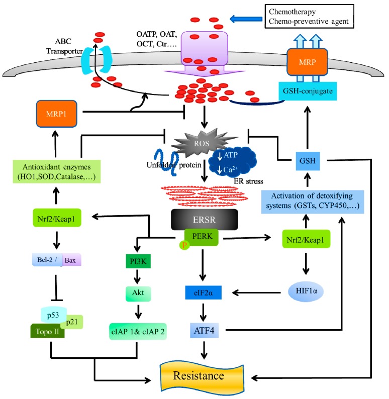
Cancers cells have the ability to develop chemotherapy resistance, which is a persistent problem during cancer treatment. Chemotherapy resistance develops through different molecular mechanisms, which lead to modification of the cancer cells signals needed for cellular proliferation or for stimulating an immune response. The endoplasmic reticulum (ER) is an important organelle involved in protein quality control, by promoting the correct folding of protein and ER-mediated degradation of unfolded or misfolded protein, namely, ER-associated degradation. Disturbances of the normal ER functions causes an accumulation of unfolded or misfolded proteins in the ER lumen, resulting in a condition called "ER stress (ERS)." ERS triggers the unfolded protein response (UPR)-also called the ERS response (ERSR)-to restore homeostasis or activate cell death. Although the ERSR is one emerging potential target for chemotherapeutics to treat cancer, it is also critical for chemotherapeutics resistance, as well. However, the detailed molecular mechanism of the relationship between the ERSR and tumor survival or drug resistance remains to be fully understood. In this review, we aim to describe the most vital molecular mechanism of the relationship between the ERSR and chemotherapy resistance. Moreover, the review also discusses the molecular mechanism of ER stress-mediated apoptosis on cancer treatments.

Keywords: cancer; chemotherapy resistance; endoplasmic reticulum; endoplasmic reticulum stress response.


Conflict of interest statement

The authors declare no conflict of interest.

Figures

Figure 1
Figure 1
Pathways involved in tumor cell death. The chemotherapeutic treatment or chemotherapy mainly utilize three different pathways that lead to tumor cell death or apoptosis, which are (a) the extrinsic death receptor pathway, (b) the intrinsic mitochondrial pathway, and (c) the endoplasmic reticulum stress (ERS) pathway.
Figure 2
Figure 2
Endoplasmic reticulum stress (ERS)-mediated apoptosis and cancer therapy. Cancers can be treated with radiotherapy or chemotherapy, either individually or as a combination therapy. The chemotherapeutic exerts its anticancer activity by inducing ERS through endoplasmic reticulum stress response (ERSR) to kill tumor cells. The ERSR is mainly transduced by three ER-resident sensor proteins, protein kinase RNA-like endoplasmic reticulum kinase (PERK), activating the transcription factor 6 alpha (ATF6), and inositol requiring enzyme 1 alpha (IRE1). The integrated signaling downstream of these three sensors can induce pro-apoptotic signaling through the activation of C/EBP homologous protein (CHOP) that can downregulate B-cell lymphoma-2 (Bcl-2) protein and upregulate Bcl-2-associated X (Bax) protein. The Ca2+ released through inositol 1,4,5-trisphosphate receptors (IP3Rs) and ryanodine receptors (RyRs) at ER and mitochondrial contact sites can promote the release of mitochondria cytochrome c which interacts with apoptosis protease-activating factor-1 (Apaf-1), adenosine triphosphate (ATP) and procaspase-9 to form the apoptosome, and subsequently activates the caspase pathway resulting in apoptosis.
Figure 3
Figure 3
ER stress and chemo-resistance: Role of protein kinase R-like endoplasmic reticulum kinase (PERK). The anti-neoplastic drugs, possess ERS-mediated apoptosis and exert their anticancer effect by entering the tumor cells, using influx transporters, including an organic anion transporter (OAT), organic anion-transporting polypeptide (OATP), organic cation transporter (OCT), and a copper transporter (Ctr). On the other hand, ATP-dependent multidrug transporters of the carrier-mediated, ubiquitous superfamily of ATP-binding cassette (ABC) proteins actively efflux chemotherapeutic agents out of the cell. ABC proteins include the multidrug resistance protein (MDR), which confer resistance to many anticancer drugs in cancer cells. The ERS-mediated activated PERK, phosphorylates eukaryotic translation initiation factor-2α (eIF2α) and nuclear factor erythroid 2 (NFE2)-related factor 2 (Nrf2). Activation of PERK via agents that trigger the ERSR, promotes a dissociation of the cytoplasmic Nrf2/Keap1, and the Nrf2 possess its cyto-protective activity by activating detoxifying enzyme systems, including the cytochrome P450 (CYP) system, glutathione-S-transferases (GSTs), thus, reducing ROS, leading to the prevention of apoptosis in cancer cells. The GSTs ensure a high GSH concentration and GSH-transporters inside cancer cells, which are associated with a high resistance to chemotherapeutic agents. The GSH exhibits cancer cell resistance to anti-neoplastic treatments, either by inhibition of apoptosis or chemotherapeutic drug detoxification, by the GSH-drug conjugation, followed by efflux through the GSH-pump of MRP transporters. The activated PERK-Nrf2 signaling, upregulates the multidrug resistance protein 1 (MRP1), by the activation of antioxidant enzymes (HO-1, SOD, catalase, etc.) that neutralize the reactive oxygen species (ROS) and increases the drug efflux, leading to a reduction in the ROS levels. Nrf2 stabilization by the exposure of antioxidant enzymes, leads to an increased Bcl-2/Bax ratio, subsequently reducing apoptosis and increasing cell survival, by regulation of Topo II, p53, and its target gene p21. The PERK activity can induce cellular inhibitor of apoptosis (cIAP1 and cIAP2) proteins, by the activation of phosphatidylinositol 3-kinase (PI3K)-Akt signaling pathway, leading to chemotherapy resistance.
Figure 4
Figure 4
ER stress and chemo-resistance: Role of inositol-requiring enzyme 1 alpha (IRE1), activating transcription factor 6 alpha (ATF6), and glucose-regulated protein 78 (GRP78). Upon ERS, IRE splices and activates the X-box-binding protein 1 (XBP1) that controls cycline A1 expression and regulates HIF1α targets. The activation of IRE1 leads to the activation of c-Jun N-terminal protein kinase (JNK), followed by inducing AP-1 transcriptional activity. The Raf/MEK/ERK pathway is also involved in IRE1-mediated chemo-resistance. ATF6α possesses chemo-resistance through the regulation of ER chaperone BiP protein, mediated by the activation of disulfide isomerase A5 (PDIA5) and p38-dependent ATF6α-mTOR pathway, upon ER stress. GRP78 exhibits its chemo-resistance properties, upon ERS, through the suppression of C/EBP homologous protein (CHOP), Bcl-2-interacting killer (BIK), phorbol-12-myristate-13-acetate-induced protein 1 (PMAIP1) or NOXA, Topo II, and induction of Bcl-2, leading to the activation of caspase-7 dependent apoptosis. GRP78 suppresses G1/S transition-related cyclins (D1, E1, and E2) and cyclin-dependent kinase (CDK4 and CDK6) protein expression. Additionally, the phosphorylation of GRP78 leads to the activation of Ras/MAPK and PI3-kinase downstream signaling, and the recruitment of p21 activated kinases 2 (PAK-2) via NCK, thus, inhibiting the pro-apoptotic protein Bcl-2-associated death (Bad).

References

    1. Global Burden of Disease Cancer Collaboration. Fitzmaurice C., Dicker D., Pain A., Hamavid H., Moradi-Lakeh M., MacIntyre M.F., Allen C., Hansen G., Woodbrook R., et al. The Global Burden of Cancer 2013. JAMA Oncol. 2015;1:505–527. - PMC - PubMed
    1. Heron M., Anderson R.N. Changes in the Leading Cause of Death: Recent Patterns in Heart Disease and Cancer Mortality. NCHS; Hyattsville, MD, USA: 2016. pp. 1–8. NCHS Data Brief 2016. - PubMed
    1. Korean Statistical Information Service National Cancer Registration Data. [(accessed on 6 July 2018)]; Available online: www.kosis.kr.
    1. Chakraborty S., Rahman T. The difficulties in cancer treatment. Ecancermedicalscience. 2012;6:ed16. - PMC - PubMed
    1. Liu B., Ezeogu L., Zellmer L., Yu B., Xu N., Joshua Liao D. Protecting the normal in order to better kill the cancer. Cancer Med. 2015;4:1394–1403. doi: 10.1002/cam4.488. - DOI - PMC - PubMed

LinkOut - more resources