Long non-coding RNA CASC2 suppresses pulmonary artery smooth muscle cell proliferation and phenotypic switch in hypoxia-induced pulmonary hypertension
- PMID: 30857524
- PMCID: PMC6413462
- DOI: 10.1186/s12931-019-1018-x
Long non-coding RNA CASC2 suppresses pulmonary artery smooth muscle cell proliferation and phenotypic switch in hypoxia-induced pulmonary hypertension
Abstract
Background: In this study, we aimed to investigate whether and how lncRNA CASC2 was involved in hypoxia-induced pulmonary hypertension (PH)-related vascular remodeling.
Methods: The expression of lncRNAs or mRNAs was detected by qRT-PCR, and western blot analysis or immunochemistry was employed for detecting the protein expression. Cell number assay and EdU (5-ethynyl-2'-deoxyuridine) staining were performed to assess cell proliferation. Besides, flow cytometry and wound healing assay were employed for assessments of cell apoptosis and cell migration, respectively. Rat model of hypoxic PH was established and the hemodynamic measurements were performed. Hematoxylin and eosin (HE) and Masson's trichrome staining were carried out for pulmonary artery morphometric analysis.
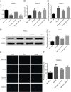
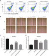
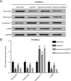
Results: The expression of lncRNA CASC2 was decreased in hypoxia-induced rat pulmonary arterial tissues and pulmonary artery smooth muscle cells (PASMCs). Up-regulation of lncRNA CASC2 inhibited cell proliferation, migration yet enhanced apoptosis in vitro and in vivo in hypoxia-induced PH. Western blot analysis and immunochemistry showed that up-regulation of lncRNA CASC2 greatly decreased the expression of phenotype switch-related marker α-SMA in hypoxia-induced PH. Furthermore, it was indicated by the pulmonary artery morphometric analysis that lncRNA CASC2 suppressed vascular remodeling of hypoxia-induced rat pulmonary arterial tissues.
Conclusion: LncRNA CASC2 inhibited cell proliferation, migration and phenotypic switch of PASMCs to inhibit the vascular remodeling in hypoxia-induced PH.
Keywords: Pulmonary artery smooth muscle cells (PASMCs); Pulmonary hypertension; Vascular remodeling; phenotypic switch; lncRNA CASC2.
Conflict of interest statement
Ethics approval and consent to participate
This study was authorized by the Fuwai Hospital, and obtained written informed consents from all the participants.,
Consent for publication
Not applicable.
Competing interests
The authors declare that they have no competing interests.
Publisher’s Note
Springer Nature remains neutral with regard to jurisdictional claims in published maps and institutional affiliations.
Figures


Similar articles
-
LncRNA CASC2 inhibits hypoxia-induced pulmonary artery smooth muscle cell proliferation and migration by regulating the miR-222/ING5 axis.Cell Mol Biol Lett. 2020 Mar 17;25:21. doi: 10.1186/s11658-020-00215-y. eCollection 2020. Cell Mol Biol Lett. 2020. PMID: 32206065 Free PMC article.
-
LncRNA-TCONS_00034812 in cell proliferation and apoptosis of pulmonary artery smooth muscle cells and its mechanism.J Cell Physiol. 2018 Jun;233(6):4801-4814. doi: 10.1002/jcp.26279. Epub 2018 Jan 15. J Cell Physiol. 2018. PMID: 29150946
-
LncRNA-SMILR modulates RhoA/ROCK signaling by targeting miR-141 to regulate vascular remodeling in pulmonary arterial hypertension.Am J Physiol Heart Circ Physiol. 2020 Aug 1;319(2):H377-H391. doi: 10.1152/ajpheart.00717.2019. Epub 2020 Jun 19. Am J Physiol Heart Circ Physiol. 2020. PMID: 32559140
-
Research progress on the mechanism of phenotypic transformation of pulmonary artery smooth muscle cells induced by hypoxia.Zhejiang Da Xue Xue Bao Yi Xue Ban. 2022 Dec 25;51(6):750-757. doi: 10.3724/zdxbyxb-2022-0282. Zhejiang Da Xue Xue Bao Yi Xue Ban. 2022. PMID: 36915980 Free PMC article. Review. English.
-
Role of macrophage migration inhibitory factor in the proliferation of smooth muscle cell in pulmonary hypertension.Mediators Inflamm. 2012;2012:840737. doi: 10.1155/2012/840737. Epub 2012 Jan 18. Mediators Inflamm. 2012. PMID: 22363104 Free PMC article. Review.
Cited by
-
Epigenetic Targets for Oligonucleotide Therapies of Pulmonary Arterial Hypertension.Int J Mol Sci. 2020 Dec 3;21(23):9222. doi: 10.3390/ijms21239222. Int J Mol Sci. 2020. PMID: 33287230 Free PMC article. Review.
-
Whole transcriptome landscape in HAPE under the stress of environment at high altitudes: new insights into the mechanisms of hypobaric hypoxia tolerance.Front Immunol. 2024 Sep 12;15:1444666. doi: 10.3389/fimmu.2024.1444666. eCollection 2024. Front Immunol. 2024. PMID: 39328420 Free PMC article.
-
Unraveling the epigenetic landscape of pulmonary arterial hypertension: implications for personalized medicine development.J Transl Med. 2023 Jul 17;21(1):477. doi: 10.1186/s12967-023-04339-5. J Transl Med. 2023. PMID: 37461108 Free PMC article. Review.
-
Profiling and Molecular Mechanism Analysis of Long Non-Coding RNAs and mRNAs in Pulmonary Arterial Hypertension Rat Models.Front Pharmacol. 2021 Jun 29;12:709816. doi: 10.3389/fphar.2021.709816. eCollection 2021. Front Pharmacol. 2021. PMID: 34267668 Free PMC article.
-
Emerging role of long non-coding RNAs in pulmonary hypertension and their molecular mechanisms (Review).Exp Ther Med. 2020 Dec;20(6):164. doi: 10.3892/etm.2020.9293. Epub 2020 Oct 9. Exp Ther Med. 2020. PMID: 33093902 Free PMC article. Review.
References
MeSH terms
Substances
LinkOut - more resources
Full Text Sources
Medical

