. 2019 Mar 11;12(1):22.
doi: 10.1186/s13048-019-0496-2.

The ovarian germinal reserve and apoptosis-related proteins in the infant and adolescent human ovary

Affiliations

The ovarian germinal reserve and apoptosis-related proteins in the infant and adolescent human ovary

María Itatí Albamonte et al. J Ovarian Res. .

Abstract

Background: Normal pubertal ovary displays all stages of follicular development and a biased BAX/BCL2 protein ratio in favor of pro-apoptotic BAX protein comparable to the adult ovary. However, adolescents suffering malignant extra-gonadal disease show a limited follicle development after cytotoxic drug treatment and a reduced capacity of in vitro follicle growth. We evaluated the expression of pro- and anti-apoptotic members of the BCL2 gene family, the FAS/FAS-L proteins from the extrinsic apoptosis pathway, the germ-cell-specific marker VASA, the pluripotency marker OCT3/4, and markers of early and late apoptosis in the ovary of pubertal patients with malignant extra-gonadal disease, which received or not pre-surgery chemotherapy, entering a cryopreservation program.

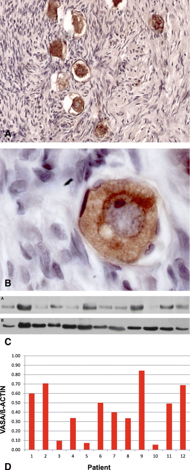
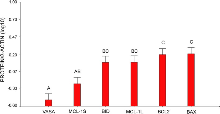
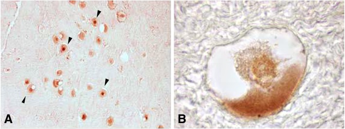
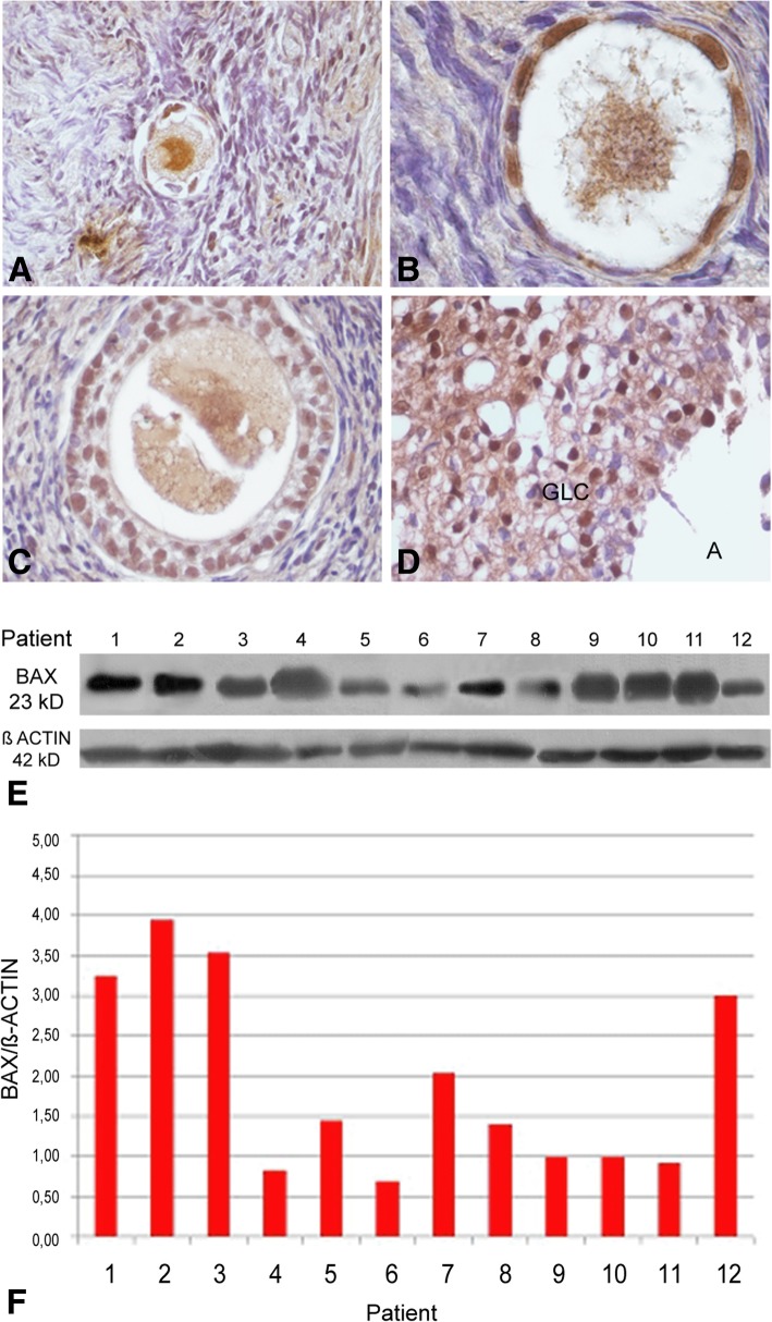
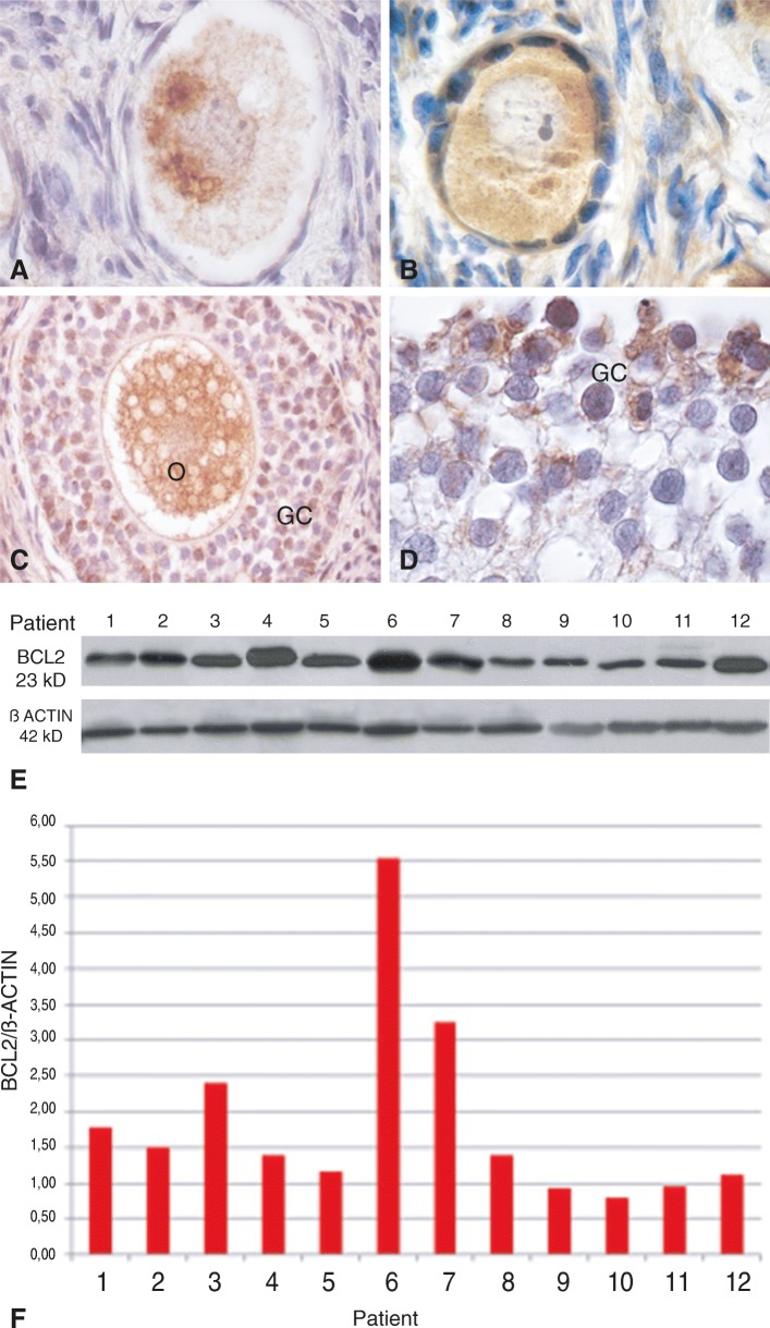
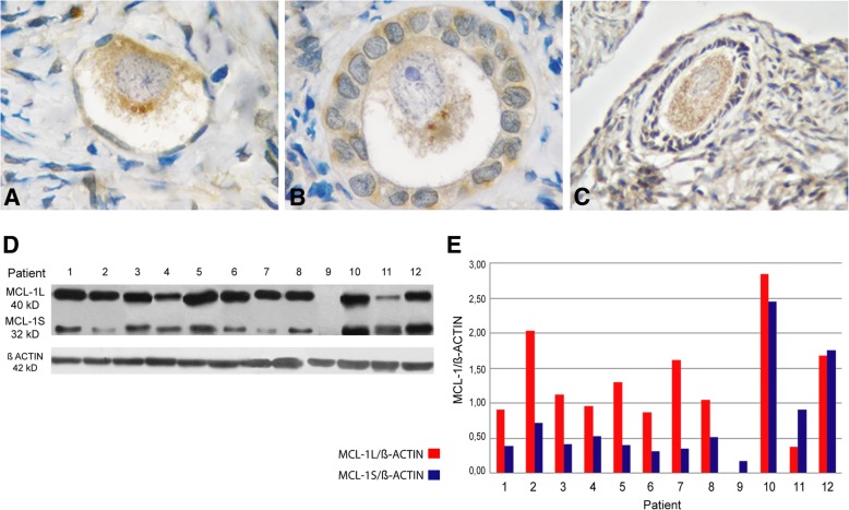
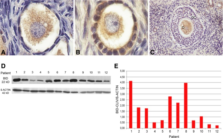
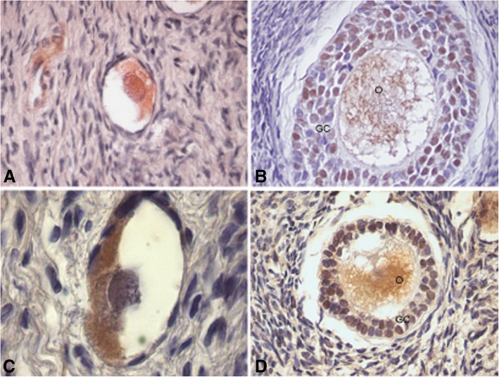
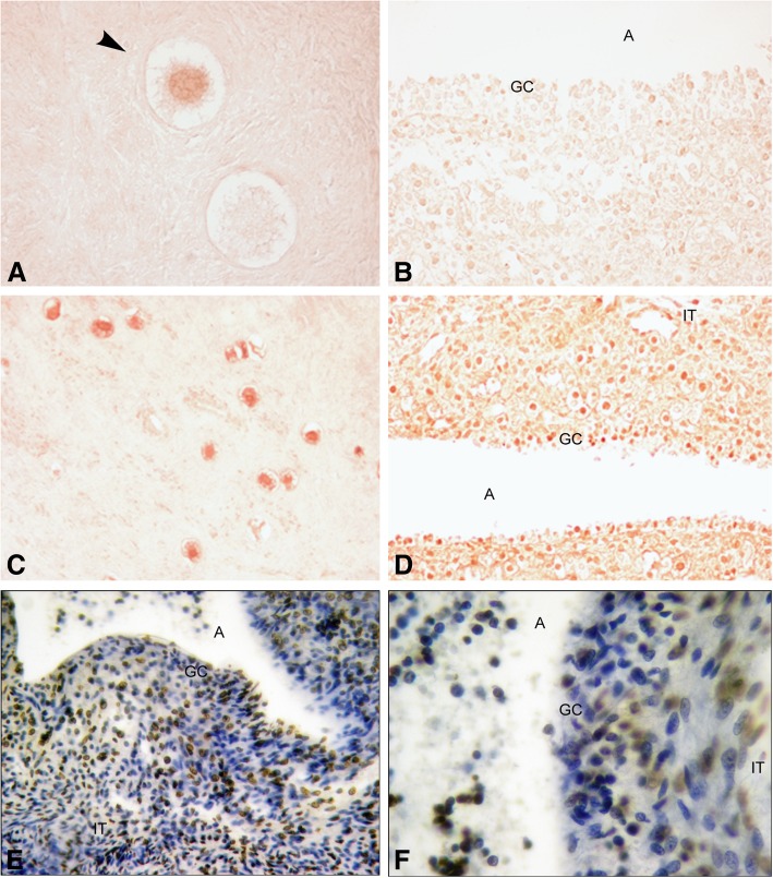
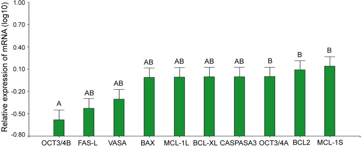
Results: Ovarian biopsies from 12 adolescent girls were screened for follicle count and expression of VASA, OCT3/4, BAX, BCL2, MCL1L and S, cleaved-BID, FAS/FAS-L and CASPASE 3 through immunohistochemistry, western blot and RT-PCR. All stages of folliculogenesis, from primordial to antral follicle, were present in all 12 patients analyzed. VASA and most of the screened apoptosis-related genes showed a pattern of immune-expression comparable to that previously reported. OCT3/4 showed a cytoplasmic localization in the great majority of the primordial follicles; however, in some cases the localization was nuclear. In addition, OCT3/4B showed a significant reduction compared to OCT3/4A. Unexpectedly, BCL2 was detected at all stages of folliculogenesis, associated to the Balbiani's body in the primordial follicles, regardless of whether patients had or had not received chemotherapy, ruling out the possibility that its expression is a protective response to chemotherapy.

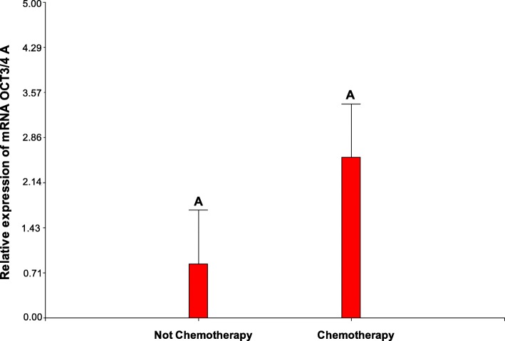
Conclusions: These findings reveal new information on the morphological status of the follicular reserve and the expression of apoptosis-related genes in histologically normal adolescent ovary from patients undergoing extragonadal cancer. The unexpected expression of apoptosis-inhibiting BCL2 protein, both in patients that had or had not received chemotherapy, opens a new avenue for thorough investigations. Moreover, the nuclear localization of OCT3/4 protein in primordial follicle-enclosed oocytes suggests a possible increased activity of ovarian stem cells in response to chemotherapy and/or extragonadal cancer. This new information can be essential for a better managing of in vitro culture of follicles that can be removed by filtration from preserved ovarian tissue, especially in girls that entered a cryopreservation program.

Keywords: Adolescence; Apoptosis; BCL2-family proteins; FAS-FAS-L; Human ovary; OCT3/4; Ovarian reserve; VASA.


Conflict of interest statement

Ethics approval

All procedures performed in this study were approved by the institutional committees from Universidad Maimónides and from Hospital de Niños Ricardo Gutiérrez, Buenos Aires, Argentina. Informed Consent was obtained from all individual participants, and/or their legal representatives, included in this study. The study followed the ethical standards from the 1964 Helsinki declaration and its later amendments.

Consent for publication

Not applicable.

Competing interests

The authors declare that they have no competing interests.

Publisher’s Note

Springer Nature remains neutral with regard to jurisdictional claims in published maps and institutional affiliations.

Figures

Fig. 1
Fig. 1
Detection of germ-cell-specific VASA protein in human ovary. a Primordial follicles VASA positive in cytoplasm oocyte (200X). b Primordial follicle VASA positive with para-nuclear localization corresponding to Balbiani’s vitelline body (1000X). c) Western blot analysis of human ovarian VASA protein (79 kD). d Western blot quantification of VASA protein. This protein was detectable in all samples with variable intensity
Fig. 2
Fig. 2
Western blot statistical analysis for VASA, BAX, BCL2, MCL-1 and cleaved-BID proteins in human ovary from pre- and pubertal oncological patients. Each bar represents a mean ± s.d. Different letters over the bars indicate significant differences between samples (Tukey, α: 0,05, p < α)
Fig. 3
Fig. 3
Immune-localization of OCT3/4 protein in adolescent human ovarian tissue. a Primordial follicles OCT3/4 positive. Note some follicles were positive in oocyte cytoplasm and others were positive in nucleus (Arrows head) (100X). b Detailed view of a typical primordial follicle; OCT3/4 protein was detectable in the cytoplasm of oocyte (1000X)
Fig. 4
Fig. 4
Detection of pro-apoptotic BAX protein in adolescent human ovary. a Primordial follicles show BAX protein in granulosa cells and in the germ-cell proper (200X). b The signal persists in primary follicle (1000X) and c) secondary follicle (400X). d Partial view of a luteum body positive for BAX (400X). e Western blot analysis of human ovarian BAX protein (23 kD). f Western blot quantification of BAX protein. This protein was detectable in all samples. GLC: granulosa-luteal cells
Fig. 5
Fig. 5
Detection of anti-apoptotic BCL2 protein in adolescent human ovary. a Primordial follicle and b) primordial/primary follicle BCL2 positive. BCL2 was heterogeneously distributed in a para-nuclear localization in the oocyte cytoplasm (Arrow heads) (1000X). c Secondary follicles show BCL2 protein in granulosa cells and in the germ-cell proper (200X). d Antral follicle (partial view), BCL2 is detectable in granulosa cells (1000X). e Western blot analysis of human ovarian BCL2 protein (23 kD). f Western blot quantification of BCL2 protein. It was detectable in all samples with variable intensity. O: oocyte; GC: granulosa cells
Fig. 6
Fig. 6
Detection of MCL-1 protein in adolescent human ovary. a Primordial follicle (1000X), b primary follicle (1000X) and c) secondary follicle (200X) show MCL-1 protein in granulosa cells and in the germ-cell proper. d Western blot analysis for both isoforms of MCL-1, the anti-apoptotic MCL-1 L (40 kD) and the pro-apoptotic MCL-1S (32 kD). e Western blot quantification of MCL-1 L and MCL-1S proteins. Both isoforms were detectable in all samples with variable intensity
Fig. 7
Fig. 7
Detection of pro-apoptotic cleaved-BID protein in adolescent human ovary. a Primordial follicle and b) primary follicle cleaved-BID positive (1000X). c Secondary follicles show cleaved-BID protein in granulosa cells and in the germ-cell proper (200X). d Western blot analysis of human ovarian cleaved-BID protein (22 kD). It was detectable in all samples. e Western blot quantification of cleaved-BID protein. It was detectable in all samples with variable intensity. O: oocyte; GC: granulosa cells
Fig. 8
Fig. 8
Immunostaining for both FAS/FAS-L proteins in adolescent human ovary. a Primary follicle (400X) and b) secondary follicle (400X) were positive for FAS protein. In the last one, it was detectable in oocyte and granulosa cells. c Primordial follicle positive for FAS-L in cytoplasm oocyte (1000X). d Secondary follicle positive for FAS-L in oocyte and granulose cells (400X). O: oocyte; GC: granulosa cells
Fig. 9
Fig. 9
Immunodetection for CASPASA-3 and TUNEL-assayed in human ovary. a Primordial follicles positive (arrow head) and negative for pro-caspase 3 (400X). b Partial view of an atretic antral follicle negative for pro-caspase 3 (200X). c Panoramic view of cortex ovary showing abundance of primordial follicles positive for cleaved caspase 3 protein (100X). d Atretic antral follicle was positive for cleaved caspase 3 in granulosa and thecal cells (200X). e Apoptotic cells were detected in granulosa and thecal cells of antral follicles (400X). f Partial view of an atretic antral follicle with granulosa cells TUNEL positive (1000X). A: antrum; GC: granulosa cells, IT: internal theca
Fig. 10
Fig. 10
mRNA expression in human ovary from pre- and pubertal oncological patients. Each bar represents a mean ± s.d. Different letters over the bars indicate significant differences between samples (Tukey, α: 0,05, p < α)
Fig. 11
Fig. 11
OCT3/4-A gene activity in patients that had or had not received chemotherapy. No significant statistical differences were detected between groups; however, gene activity tends to be increased in response to chemotherapy

Similar articles

Cited by

References

    1. Aitken RJ, Findlay JK, Hutt KJ, Kerr JB. Apoptosis in the germ line. Reproduction. 2011;141:139–150. doi: 10.1530/REP-10-0232. - DOI - PubMed
    1. Albamonte MI, Albamonte MS, Stella I, Zuccardi L, Vitullo AD. The infant and pubertal human ovary: Balbiani’s body associated VASA expression, immunohistochemical detection of apoptosis-related BCL2 and BAX proteins, and DNA fragmentation. Hum Reprod. 2013;28:698–706. doi: 10.1093/humrep/des453. - DOI - PubMed
    1. Albamonte MS, Albamonte MI, Vitullo AD. Germ line apoptosis in the mature human ovary. J Med Res Sci. 2012;2:136–145.
    1. Albamonte MS, Willis MA, Albamonte MI, Jensen F, Espinosa MB, Vitullo AD. The developing human ovary: immunohistochemical analysis of the germ cell-specific VASA protein, BCL-2/BAX expression balance and apoptosis. Hum Reprod. 2008;23:1895–1901. doi: 10.1093/humrep/den197. - DOI - PubMed
    1. Anderson R, Fulton N, Cowan G, Coutts S, Saunders P. Conserved and divergent patterns of expression of DAZL, VASA and OCT4 in the germ cells of the human fetal ovary and testis. BMC Develop Biol. 2007;7:136. doi: 10.1186/1471-213X-7-136. - DOI - PMC - PubMed

Substances