

A modified preauricular and transmandibular approach for surgical management of osteosarcoma of the mandibular condyle within the masticator space and infratemporal fossa: a case report
- PMID: 30857556
- PMCID: PMC6413457
- DOI: 10.1186/s13256-019-1975-1
A modified preauricular and transmandibular approach for surgical management of osteosarcoma of the mandibular condyle within the masticator space and infratemporal fossa: a case report
Abstract
Background: Osteosarcomas of the head and neck region are rare entities that comprise < 10% of all osteosarcomas. Multimodality treatment of patients with osteosarcoma is well-established for osteosarcoma in long bones, and the benefits of chemotherapy in long bones are clearly known. However, there is no consensus regarding the effects of chemotherapy in cases of head and neck osteosarcoma. The prognostic factor for head and neck osteosarcoma is complete tumor resection with negative margin, which is a radical surgery. However, a clear margin may be difficult to achieve in the head and neck region.
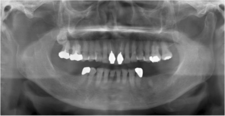
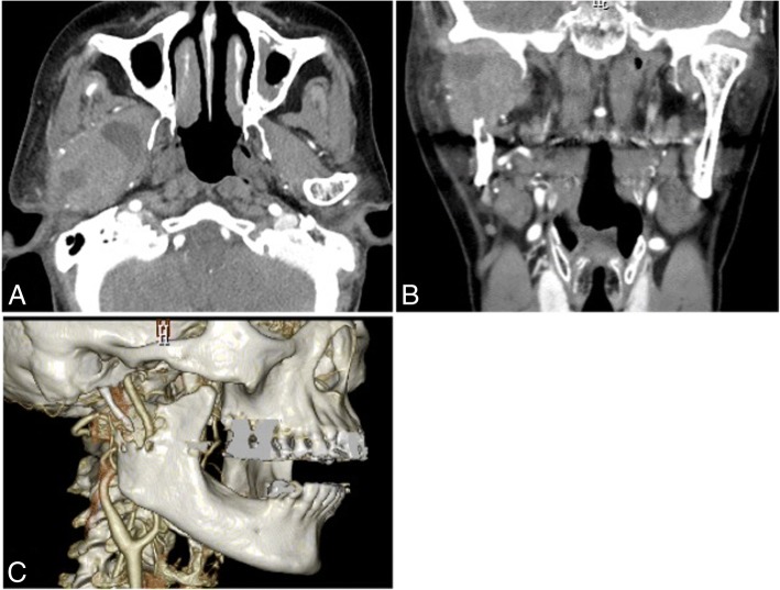
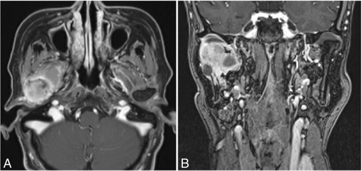
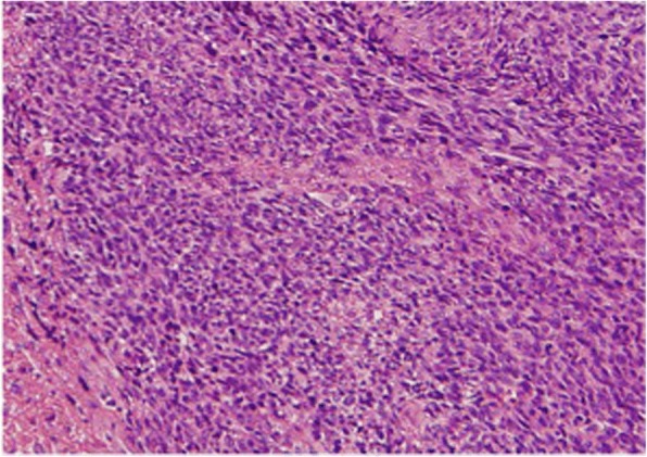
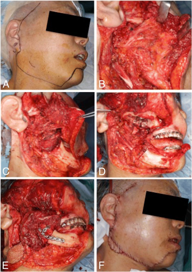
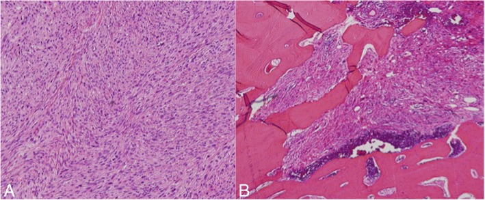
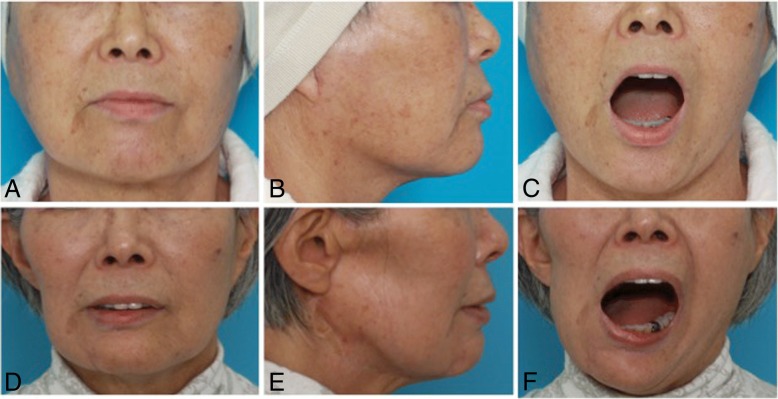
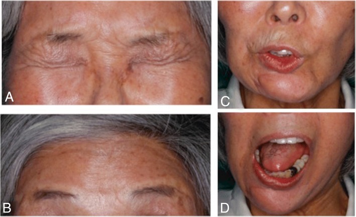
Case presentation: We present a case of a 69-year-old Japanese woman who developed osteosarcoma of the condyle within the masticator space and infratemporal fossa, which was treated with radical surgery using a modified preauricular and transmandibular approach. Although we recommended adjuvant treatment after surgery, the patient refused this treatment. There was no evidence of local recurrence or distant metastasis through 30 months of follow-up.
Conclusions: Our modified preauricular and transmandibular approach allowed access to the masticator space and infratemporal fossa, thereby increasing complete resection of the tumor and resulting in minimal functional and cosmetic deficits.
Keywords: Mandibular condyle; Masticator space; Osteosarcoma; Preauricular approach; Transmandibular approach.
Conflict of interest statement
Ethics approval and consent to participate
Not applicable.
Consent for publication
Written informed consent was obtained from the patient for publication of this case report and any accompanying images. A copy of the written consent is available for review by the Editor-in-Chief of this journal.
Competing interests
The authors declare that they have no competing interests.
Publisher’s Note
Springer Nature remains neutral with regard to jurisdictional claims in published maps and institutional affiliations.
Figures
References
-
- Cohen IJ. Significant recent advances in the treatment of osteosarcoma. Isr J Med Sci. 1993;29:748–753. - PubMed
-
- Ajura AJ, Lau SH. A retrospective clinicopathologocal study of 59 osteogenic sarcoma of jaw bone archived in a stomatology unit. Malays J Pathol. 2010;32:27–34. - PubMed
-
- Rosen G, Capparros B, Huvos AG, Kosloff C, Nirenberg A, Cacavio A, et al. Preoperative chemotherapy for osteogenic sarcoma: selection of postoperative adjuvant chemotherapy based on the response of the primary tumor preoperative chemotherapy. Cancer. 1982;49:1221–1230. doi: 10.1002/1097-0142(19820315)49:6<1221::AID-CNCR2820490625>3.0.CO;2-E. - DOI - PubMed
Publication types
MeSH terms
LinkOut - more resources
Full Text Sources
