. 2019 Mar 11;7(1):38.
doi: 10.1186/s40478-019-0693-7.

Immunopathological characterization of ovarian teratomas associated with anti-N-methyl-D-aspartate receptor encephalitis

Affiliations

Immunopathological characterization of ovarian teratomas associated with anti-N-methyl-D-aspartate receptor encephalitis

Aude Chefdeville et al. Acta Neuropathol Commun. .

Abstract

Encephalitis with anti-NMDAR antibodies (NMDAR-E) is a severe autoimmune neurological disorder, defined by a clinical presentation of encephalitis and the presence of IgG targeting the GluN1 subunit of NMDA receptors in the CSF. An underlying ovarian teratoma is commonly associated with this autoimmune disease suggesting a role of the tumor in immunopathogenesis. In this study, we characterized the salient histopathological features of 27 ovarian teratomas associated with NMDAR-E (3 immature and 24 mature teratomas) and 40 controls without associated encephalitis. All but one NMDAR-E-associated teratomas contained a nervous tissue component, while less than 40% of control teratomas did (p < 0.001). GluN1 expression by teratomatous nervous tissue seemed to be more often glial in NMDAR-E teratomas than in control teratomas (73% vs. 29%, p < 0.05). Strikingly, 3 out of 24 NMDAR-E-associated mature teratomas contained neuroglial tissue exhibiting histopathological features of central nervous system neuroglial tumor, while such glioma-like features are exceptionally described in the literature on ovarian teratomas. Moreover, NMDAR-E associated teratomas differed from sporadic ovarian teratomas by consistent and prominent infiltration of the nervous tissue component by immune cells, comprised of T- and B-cells and mature dendritic cells organized in tertiary lymphoid structures, with IgG and IgA deposits and plasma cells in close contact to the neuroglial tissue.These data demonstrate an association between massive infiltration of NMDAR-E-associated teratomas by immune cells and particular glial features of its neuroglial component, suggesting that this glial tissue might be involved in triggering or sustaining the anti-tumor response associated with the auto-immune neurological disease.

Keywords: Anti-NMDAR encephalitis; Autoimmunity; Ovarian teratoma.


Conflict of interest statement

Ethics approval and consent to participate

Written consent was obtained from all patients, and this study was approved by the institutional review board of the University Claude Bernard Lyon 1 and Hospices Civils de Lyon.

Competing interests

The authors have declared that they have no competing interests.

Publisher’s Note

Springer Nature remains neutral with regard to jurisdictional claims in published maps and institutional affiliations.

Figures

Fig. 1
Fig. 1
Central nervous tissue in NMDAR-E-associated and control ovarian teratomas. (a-b) Representative hematoxylin-phloxine-saffron (HPS) gross structure of NMDAR-E mature ovarian teratomas. In case (a), a strip of central nervous tissue (outlined, black-dotted lines) lined the inner wall of a cystic cavity. In case (b), the central nervous tissue (outlined, black-dotted lines) formed a solid mass between connective and adipose tissues (c-d) Representative HPS structure of immature ovarian teratomas associated with NMDAR-E. In (c), note the strip of central nervous tissue (outlined, black dotted lines) and the immature foci (arrowheads) with increased cellular density and neuroepithelial tubules. In case (d), the immature nervous contingent formed a solid mass lined with ependymal wall; note the choroid plexuses (arrowhead). e-f Representative HPS structure of control mature teratomas. The case (e) contained a strip of mature nervous tissue lining the wall of a cystic cavity (outlined, black dotted line). In the case (f), the solid mature nervous tissue was surrounded by connective and adipose tissue. Scale bar: 100 μm
Fig. 2
Fig. 2
GluN1 expression by neuronal and glial cells in NMDAR-E associated and control ovarian teratomas. a Representative neuronal GluN1 immunostaining of ovarian teratomas associated with NMDAR-E. Neuronal expression of NMDAR subunit is detected in large ganglion cells (see cytoplasmic and weak nuclear staining on magnification). b Representative neuronal GluN1 immunostaining in the nervous tissue of a sporadic ovarian teratoma. Note the cerebellar-like organization with Purkinje-like cells expressing GluN1 (on magnification). c Representative glial GluN1 immunostaining of ovarian teratomas associated with NMDAR-E. GluN1 expression by astrocytic cells is cytoplasmic but also sometimes nuclear (on magnification). d Representative GluN1 immunostaining of sporadic mature ovarian teratomas with a nervous component. NMDAR subunit was expressed by neuropil, cell bodies of ganglion cells (arrows) and glial cells (on magnification). e-g Representative co-immunofluorescence stainings for GFAP (green) and GluN1 (red) in the central nervous tissue of NMDAR-E teratomas. h-j Representative co-immunofluorescence stainings for GFAP (green) and GluN1 (red) in the central nervous tissue of control teratomas. Scale bar: 50 μm
Fig. 3
Fig. 3
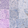
Histopathological phenotypes of NMDAR-E associated teratoma presenting histological features of gliomas. a-c Case #4. Hematoxylin-phloxine-saffron (HPS) staining (a) of the solid nervous tissue containing monotonous cells surrounded by perinuclear cytoplasmic halos (“fried-egg” oligodendrocytes-like cells) mixed with a vascular network consisting of short geometrically-arranged capillary segments, consistent with the histopathological observations of an oligodendroglioma. Some tumor cells showed positivity for Olig2 expression (b) but low Ki-67 proliferation index (c). d-f Case #5. HPS staining (d) of the neuroglial tissue showing increased cellular density, clusters of ganglion cells and aligned oligodendrocytes-like cells, consistent with the histological features of a ganglioglioma. Some tumor cells were found to be positive for Olig2 expression (e) and the Ki-67 proliferation index was increased in the perivascular area (f). g-i Case #6. HPS staining (g) of the nervous tissue with highly elevated cellular density and pleiomorphic or poorly differentiated cells, consistent with the histopathological aspect of a malignant glioma. Some tumor cells show positivity for Olig2 expression (h) and elevated Ki-67 proliferation index (i). Scale bar: 100 μm
Fig. 4
Fig. 4
Inflammatory infiltrates in contact with nervous tissue in NMDAR-E-associated and control ovarian teratomas. Representative hematoxylin-phloxine-saffron (HPS) staining of teratoma nervous tissue with no (score 0), weak (score 1+), moderate (score 2+) or high (score 3+) immune cell infiltration (dotted). Scale bars: 50 μmn. Comparison of the semi-quantitative analysis of immune cell infiltrates between NMDAR-E associated (n = 26) and control (n = 15) ovarian teratomas with nervous tissue. Scale bar: 50 μm
Fig. 5
Fig. 5
Characterization of the immune environment of the nervous tissue of NMDAR-E-associated ovarian teratomas. Representative nervous tissue of NMDAR-E teratoma infiltrated by immune cells and stained by hematoxylin-phloxine-saffron (a, f, k) or immunolabeled with the neuronal marker NF (b, g, l), the CD3 T-cell marker (c, h, m), the CD20 B-cell marker (d, i, n), or the mature dendritic cell marker DC-LAMP (e, j, o). a-e Diffuse inflammatory infiltrate closely adjacent to neural tissue (a, dotted area) containing NF-stained neurons (b) and composed of T-cells (c) and B-cells (d), with some mature dendritic cells (e, see higher magnification in the upper right corner). (f-j) Tertiary Lymphoid Structure (TLS) next to a focus of neuroglial tissue (f, dotted area) containing some NF positive neurites (g), with segregated B- and T-cells (h, i) and mature dendritic cells (j, see higher magnification in the upper right corner). k-o Lymphoid infiltrates close to a foci of neuroglial tissue (k, dotted area) without NF expression (l) and composed of segregated T-cells (m) and B-cells (n). Note the absence of mature dendritic cells (o, see higher magnification in the upper right corner). Scale bars: 50 μm
Fig. 6
Fig. 6
IgG and IgA deposits and producing cells in NMDAR-E associated teratomas. a Representative GFAP/IgG/IgA immunofluorescence (IF) staining of a NMDAR-E associated teratoma showing IgG (in red) and IgA (in white) deposits along a GFAP+ strip of neuroglial tissue (in green). Hoechst was used to visualize nuclei (blue) and pictures on the right correspond to individual channels. b Representative GFAP/IgG/IgA IF staining of a control teratoma without IgG nor IgA deposits in contact with GFAP+ neuroglial tissue. c GFAP/IgG/IgA IF staining of a NMDAR-E associated teratoma focused on GFAP+ neuroglial islands surrounded by immune cells and diffuse IgA deposits (white) and some isolated IgG+ cells (red). Hoechst was used to visualize nuclei (blue) and pictures on the right correspond to individual channels. d GFAP/IgG/IgA IF staining of a NMDAR-E teratoma showing individualized IgA+ cells (white) and IgG+ (red) in contact with GFAP+ neural elements (green) in a NMDAR-E associated teratoma. Scale bars: 50 μm

Similar articles

Cited by

References

    1. Berger N, Pochaczevsky R. Astrocytoma-containing ovarian teratoma in childhood. Am J Roentgenol Radium Therapy, Nucl Med. 1969;107:647–651. doi: 10.2214/ajr.107.3.647. - DOI - PubMed
    1. Bost C, Chanson E, Picard G, Meyronet D, Mayeur M-E, Ducray F, Rogemond V, Psimaras D, Antoine J-C, Delattre J-Y, Desestret V, Honnorat J (2018) Malignant tumors in autoimmune encephalitis with anti-NMDA receptor antibodies. J Neurol. 10.1007/s00415-018-8970-0 - PubMed
    1. Büyükka Bay S, Corapcioglu F, Kavurt S, Müezzinoğlu B, Anik Y, Tugay M. Oligodendroglioma arising in a mature cystic ovarian teratoma in a child. Pediatr Hematol Oncol. 2010;27:636–640. doi: 10.3109/08880018.2010.505633. - DOI - PubMed
    1. Caruso PA, Marsh MR, Minkowitz S, Karten G. An intense clinicopathologic study of 305 teratomas of the ovary. Cancer. 1971;27:343–348. doi: 10.1002/1097-0142(197102)27:2<343::AID-CNCR2820270215>3.0.CO;2-B. - DOI - PubMed
    1. Conti F, Barbaresi P, Melone M, Ducati A. Neuronal and glial localization of NR1 and NR2A/B subunits of the NMDA receptor in the human cerebral cortex. Cerebral cortex. 1999;9:110–120. doi: 10.1093/cercor/9.2.110. - DOI - PubMed

Publication types

MeSH terms

Supplementary concepts