. 2019 Apr 26;294(17):6843-6856.
doi: 10.1074/jbc.RA119.007406. Epub 2019 Mar 11.

A complex containing the O-GlcNAc transferase OGT-1 and the ubiquitin ligase EEL-1 regulates GABA neuron function

Affiliations

A complex containing the O-GlcNAc transferase OGT-1 and the ubiquitin ligase EEL-1 regulates GABA neuron function

Andrew C Giles et al. J Biol Chem. .

Abstract

Inhibitory GABAergic transmission is required for proper circuit function in the nervous system. However, our understanding of molecular mechanisms that preferentially influence GABAergic transmission, particularly presynaptic mechanisms, remains limited. We previously reported that the ubiquitin ligase EEL-1 preferentially regulates GABAergic presynaptic transmission. To further explore how EEL-1 functions, here we performed affinity purification proteomics using Caenorhabditis elegans and identified the O-GlcNAc transferase OGT-1 as an EEL-1 binding protein. This observation was intriguing, as we know little about how OGT-1 affects neuron function. Using C. elegans biochemistry, we confirmed that the OGT-1/EEL-1 complex forms in neurons in vivo and showed that the human orthologs, OGT and HUWE1, also bind in cell culture. We observed that, like EEL-1, OGT-1 is expressed in GABAergic motor neurons, localizes to GABAergic presynaptic terminals, and functions cell-autonomously to regulate GABA neuron function. Results with catalytically inactive point mutants indicated that OGT-1 glycosyltransferase activity is dispensable for GABA neuron function. Consistent with OGT-1 and EEL-1 forming a complex, genetic results using automated, behavioral pharmacology assays showed that ogt-1 and eel-1 act in parallel to regulate GABA neuron function. These findings demonstrate that OGT-1 and EEL-1 form a conserved signaling complex and function together to affect GABA neuron function.

Keywords: Caenorhabditis elegans (C. elegans); E3 ubiquitin ligase; EEL-1; GABAergic transmission; HUWE1; O-linked N-acetylglucosamine (O-GlcNAc) transferase (OGT); OGT-1; gamma-aminobutyric acid (GABA); motor neuron; neurotransmission; proteomics; synapse.


Conflict of interest statement

The authors declare that they have no conflicts of interest with the contents of this article

Figures

Figure 1.
Figure 1.
Automated behavioral assay shows that eel-1 mutants are hypersensitive to aldicarb. A, depicted is the C. elegans motor circuit composed of excitatory cholinergic and inhibitory GABAergic motor neurons. Balance of contraction and relaxation is required for normal movement (left). Application of aldicarb, an AchE inhibitor, leads to excess cholinergic transmission and paralysis (middle). Mutants with impaired GABAergic transmission, such as eel-1, are hypersensitive to aldicarb and paralyze faster (right). B, schematic showing automated aldicarb assay with MWT. 20 wells are monitored simultaneously. Shown is an image of a single well with four animals (right). MWT plots depict animal swimming over a 1-min timeframe with and without aldicarb treatment (below). C, MWT analysis of aldicarb dose response for WT animals. Shown is the mean of multiple wells for each dose (n = 5–20 wells/dose); significance was determined using two-way ANOVA (dose versus time). D, eel-1 (zu462) mutants are hypersensitive to aldicarb, and transgenic expression of EEL-1 rescues aldicarb hypersensitivity. Shown are means (n = 20 wells/genotype). Inset, mean speed (line) and speed in each well (circles) at the indicated time for each genotype. Comparisons between genotypes represent pairwise two-way ANOVAs. Comparisons in the inset represent Fisher's LSD post hoc test (see Fig. S2A for further statistical analysis). ***, p < 0.001.
Figure 2.
Figure 2.
Affinity purification proteomics from C. elegans identifies OGT-1 as a putative EEL-1 binding protein. A, schematic of eel-1 gene and eel-1 (bgg1) null allele generated by MosDel. Shown are exons (black), promoter (white), UTRs (gray), and introns (gaps connected by lines). B, schematic of EEL-1 protein with annotated domain of unknown function (DUF), ubiquitin-associated domain (UBA), and HECT ubiquitin ligase domain (HECT). Highlighted is the residue required for catalytic activity (purple). C, automated aldicarb assay showing that eel-1 (bgg1) null mutants are hypersensitive to aldicarb compared with WT. Aldicarb hypersensitivity is rescued by GS::EEL-1 but not GS::EEL-1 LD. GS::GFP is negative control. Shown are means of multiple wells for each genotype (n = 10–30 wells with 4 worms/well). The inset shows mean speed (line) and individual data points at the indicated time. D, work flow for affinity purification proteomics from C. elegans. E, example of LC-MS/MS spectrum identifying OGT-1 peptide sequence from GS::EEL-1 sample. F, comparison of total peptide spectra from four proteomic experiments for proteins identified in GS::EEL-1 sample compared with GS::GFP sample. The dashed line denotes 2× enrichment above GS::GFP negative control. G, comparison of normalized total peptide spectra from four proteomic experiments for GS::EEL-1 LD compared with GS::EEL-1. Highlighted in gold are proteins found exclusively in or enriched in EEL-1 LD. The dashed line denotes the point of equivalency between GS::EEL-1 and GS::EEL-1 LD. H, summary of EEL-1 affinity purification proteomics data. OGT-1 is identified in GS::EEL-1 and GS::EEL- LD but absent in GS::GFP. OGT-1 was identified at similar levels in GS::EEL-1 and GS::EEL-1 LD when samples were normalized to the amount of EEL-1 target. For C, comparisons between genotypes represent pairwise two-way ANOVAs. Comparisons in the inset represent Fisher's LSD post hoc test. For H, significance was determined using Student's t test. ***, p < 0.001; ns, not significant (p > 0.05).
Figure 3.
Figure 3.
OGT-1 is a conserved EEL-1 binding protein that interacts with EEL-1 in neurons. A, GFP::EEL-1 was expressed alone or with FLAG::OGT-1 in the nervous system of C. elegans. Co-IP from transgenic worm lysates shows that GFP::EEL-1 binds FLAG::OGT-1. B, co-IP showing human GFP::HUWE1 binds to human FLAG::OGT when expressed in HEK 293 cells. Shown are representative images from three or more independent experiments.
Figure 4.
Figure 4.
OGT-1 is expressed in cholinergic and GABAergic motor neurons and localizes to GABAergic presynaptic terminals. A, representative epifluorescent images showing that GFP driven by the ogt-1 promoter (Pogt-1 GFP) is broadly expressed inside and outside the nervous system. B and C, schematic and representative confocal images showing that GFP expressed using the ogt-1 promoter (Pogt-1 GFP) coexpresses with markers for cholinergic (B) and GABAergic (C) motor neurons. D, schematic and representative confocal images showing that mCherry::OGT-1 colocalizes with synaptic vesicle marker SNB-1::GFP at presynaptic terminals of GABAergic motor neurons. Shown are maximum intensity z-projection (left) and single z-slices (right) from the indicated region (blue box). All scale bars are 20 μm except for the one in the right panel of D, which is 5 μm.
Figure 5.
Figure 5.
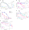
ogt-1 functions in GABA neurons to regulate aldicarb sensitivity and enhances eel-1. Shown are automated aldicarb assays with speed normalized to the buffer-only control group for each genotype. A, ogt-1 (ok430) mutants are hypersensitive to a moderate aldicarb dose (64 μm) compared with WT. Aldicarb hypersensitivity is rescued by transgenic expression of OGT-1 pan-neuronally or in GABA neurons. Shown are means of multiple wells for each genotype (n = 29–30 wells, 4 worms/well). Inset, mean speed (line) and individual data points at the indicated time. B, eel-1 (bgg1) mutants are hypersensitive to low aldicarb dose (8 μm) compared with WT, and hypersensitivity is enhanced in ogt-1; eel-1 double mutants. Shown are means (n = 10–15 wells). C, dose response showing that ogt-1 mutants are hypersensitive to moderate and high doses of aldicarb. Aldicarb hypersensitivity is enhanced in ogt-1; eel-1 double mutants across doses. Shown are mean ± S.E. for normalized speed 35 min after aldicarb exposure (n = 5–15 wells). D, OGT-1 expressed in neurons rescues enhanced aldicarb hypersensitivity of ogt-1; eel-1 double mutants. Shown are means (n = 30 wells for each genotype, 1 worm/well due to ogt-1; eel-1 low brood size). E, automated analysis of swimming speed for indicated genotypes. Swimming defects in eel-1 and ogt-1 single mutants are enhanced in ogt-1; eel-1 double mutants (n = 30–65 wells). Inset, mean speed (line) and individual data points at the indicated time. Comparisons between genotypes represent pairwise two-way ANOVAs. Comparisons in the inset represent Fisher's LSD post hoc test (see Fig. S2B for further statistical analysis). **, p < 0.01; ***, p < 0.001; ns, not significant (p > 0.05).
Figure 6.
Figure 6.
GABA synapse formation is not impaired in eel-1; ogt-1 double mutants. A, schematic of GABA NMJs in C. elegans dorsal cord. SNB-1::GFP marks GABA presynaptic terminals and GABA receptor subunit, and UNC-49::RFP marks postsynaptic terminals. B, representative confocal images showing colocalization of presynaptic SNB-1::GFP and postsynaptic UNC-49::RFP is normal for all indicated genotypes. C, quantitation indicates that the number of SNB-1::GFP puncta is not changed for any genotype. Shown are mean ± S.E. (error bars) (n = 38–44 worms). Significance was tested using ANOVA. ns, not significant (p > 0.05). Scale bar, 20 μm.
Figure 7.
Figure 7.
OGT-1 glycosyltransferase activity is not required for motor circuit function. Shown are automated aldicarb assays with speed normalized to buffer-only control group for each genotype. Aldicarb hypersensitivity in ogt-1 (ok430) mutants is significantly rescued by pan-neuronal expression of two OGT-1 point mutants that lack catalytic glycosyltransferase activity: OGT-1 K957M (A) and OGT-1 H612A (B) (n = 30 wells, 4 worms/well). C, oga-1 (ok1207) mutants have similar aldicarb hypersensitivity as WT animals. Note that ogt-1 (ok430) and rab-3 (js49) mutants are included as controls for aldicarb hypersensitivity and resistance, respectively. D, dose response showing that oga-1 mutants have similar responses to aldicarb as WT animals. Shown are mean ± S.E. (error bars) for normalized speed 45 min after aldicarb exposure (n = 20 wells). Comparisons between genotypes represent pairwise two-way ANOVAs. Comparisons in the inset represent Fisher's LSD post hoc tests. ***, p < 0.001; ns, not significant (p > 0.05).
Figure 8.
Figure 8.
OGT-1 and EEL-1 form a protein complex and regulate GABA neuron function in C. elegans motor circuit. Shown is a summary of our findings that OGT-1 and EEL-1 form a complex in vivo in C. elegans neurons. OGT-1 and EEL-1 are expressed in both cholinergic and GABAergic motor neurons but function in parallel to regulate GABA neuron function.

References

    1. Huang Z. J., and Scheiffele P. (2008) GABA and neuroligin signaling: linking synaptic activity and adhesion in inhibitory synapse development. Curr. Opin. Neurobiol. 18, 77–83 10.1016/j.conb.2008.05.008 - DOI - PMC - PubMed
    1. Krueger-Burg D., Papadopoulos T., and Brose N. (2017) Organizers of inhibitory synapses come of age. Curr. Opin. Neurobiol. 45, 66–77 10.1016/j.conb.2017.04.003 - DOI - PubMed
    1. Schuske K., Beg A. A., and Jorgensen E. M. (2004) The GABA nervous system in C. elegans. Trends Neurosci. 27, 407–414 10.1016/j.tins.2004.05.005 - DOI - PubMed
    1. Zhen M., and Samuel A. D. (2015) C. elegans locomotion: small circuits, complex functions. Curr. Opin. Neurobiol. 33, 117–126 10.1016/j.conb.2015.03.009 - DOI - PubMed
    1. Ramamoorthi K., and Lin Y. (2011) The contribution of GABAergic dysfunction to neurodevelopmental disorders. Trends Mol. Med. 17, 452–462 10.1016/j.molmed.2011.03.003 - DOI - PMC - PubMed

Publication types

MeSH terms