. 2019 May 3;294(18):7245-7258.
doi: 10.1074/jbc.RA118.005533. Epub 2019 Mar 11.

Substitutions that lock and unlock the proton-coupled folate transporter (PCFT-SLC46A1) in an inward-open conformation

Affiliations

Substitutions that lock and unlock the proton-coupled folate transporter (PCFT-SLC46A1) in an inward-open conformation

Srinivas Aluri et al. J Biol Chem. .

Abstract

The proton-coupled folate transporter (PCFT) mediates intestinal absorption of folates and their transport from blood to cerebrospinal fluid across the choroid plexus. Substitutions at Asp-109 in the first intracellular loop between the first and second transmembrane domains (TMDs) abolish PCFT function, but protein expression and trafficking to the cell membrane are retained. Here, we used site-directed mutagenesis, the substituted-cysteine accessibility method, functional analyses, and homology modeling to determine whether the D109A substitution locks PCFT in one of its conformational states. Cys-substituted residues lining the PCFT aqueous translocation pathway and accessible in WT PCFT to the membrane-impermeable cysteine-biotinylation reagent, MTSEA-biotin, lost accessibility when introduced into the D109A scaffold. Substitutions at Gly-305 located exofacially within the eighth TMD, particularly with bulky residues, when introduced into the D109A scaffold largely restored function and MTSEA-biotin accessibility to Cys-substituted residues within the pathway. Likewise, Ser-196 substitution in the fifth TMD, predicted by homology modeling to be in proximity to Gly-305, also partially restored function found in solute transporters, is critical to oscillation of the carrier among its conformational states. Substitutions at Asp-109 and Gly-112 lock PCFT in an inward-open conformation, resulting in the loss of function. However, the integrity of the locked protein is preserved, indicated by the restoration of function after insertion of a second "unlocking" mutation. and accessibility. Similarly, the inactivating G112K substitution within the first intracellular loop was partially reactivated by introducing the G305L substitution. These data indicate that the first intracellular loop, with a sequence identical to "motif A" (GXXXDXXGR(R/K)).

Keywords: GXXXDXXGR(R/K); folate; folate malabsorption disease; membrane transport; protein motif; proton-coupled folate transporter (PCFT); solute carrier family 46 member 1 (SLC46A1); structural model; transporter.


Conflict of interest statement

The authors declare that they have no conflicts of interest with the contents of this article

Figures

Figure 1.
Figure 1.
Topological structure of PCFT. The secondary structure of PCFT consists of 12 transmembrane domains with N and C termini directed to the cytoplasm (22). Residues in the first intracellular loop (Asp-109, Gly-112, and Arg-113) are highlighted in red. Cys-substituted residues studied, that are accessible in PCFT-WT, are highlighted in orange. Two residues that when mutated restore function when introduced in the D109A scaffold are highlighted in green (Gly-305 (eighth TMD) and Ser-196 (fifth TMD)). Cys-substituted residues studied in the eighth TMD are indicated in pink. Ile-188 (fifth TMD) and Leu-161 (fourth TMD) are highlighted in blue. The arrows indicate the positions of amino acids predicted to be located at membrane interfaces.
Figure 2.
Figure 2.
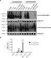
The impact of the D109A mutation on the accessibility and function of Cys-substituted mutants. A, in this Western blotting, the top row indicates Lys biotinylation at the cell membrane (left) and Cys biotinylation (right) of mutants and WT. The bottom row indicates total PCFT expression in the corresponding crude membrane extracts. Integrin β1 is the loading control for both biotinylated and crude membrane proteins. The gap between integrin β1 loading controls for Lys biotinylation and Cys biotinylation indicates different exposure times of the X-ray films. The Western blotting is representative of three independent experiments. B, [3H]MTX influx was assessed at 37 °C and pH 5.5 over 1 min at concentrations of 0.5 μm (an order of magnitude below the MTX influx Kt) and 50 μm (an order of magnitude above). Data are indicated as percentage ± S.E. (error bars) of PCFT-WT influx from three independent experiments. A one-way ANOVA compared single Cys mutants with corresponding D109A double mutants. ****, p < 0.0001.
Figure 3.
Figure 3.
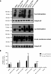
The impact of the D109A mutation on the accessibility of Cys-substituted exofacial residues within TMDs and including the region of the external gate. A, MTSEA-biotin accessibility of Cys-substituted residues with the integrin β1 loading control is shown in the top row, whereas surface expression of the mutants is indicated in the middle row. PCFT expression in the crude membrane extracts is indicated in the bottom row with the integrin β1 loading control. The Western blotting is a representative of three independent experiments. B, [3H]MTX influx was assessed at 37 °C and pH 5.5 over 1 min at concentrations of 0.5 and 50 μm. Data are represented as percentage ± S.E. (error bars) of PCFT-WT influx from three independent experiments. A one-way ANOVA compared single mutant with each double mutant. *, p < 0.05; **, p < 0.01; ****, p < 0.0001.
Figure 4.
Figure 4.
The impact of the D109A mutation on accessibility to MTSEA-biotin of resides within the exofacial cleft of the eighth TMD. A, in the top row, the accessibility of Cys-substituted residues to MTSEA-biotin of the single mutants, or in combination with the D109A mutation, are shown going from left to right in the figure from a residue (G305C) high in the exofacial region to a residue (P314C) deep within the aqueous pathway. Integrin β1 is the loading control. The middle row indicates expression at the plasma membrane. The bottom row indicates PCFT expression in the crude membrane extract with the integrin β1 loading control. The Western blotting is a representative of three independent experiments. B, [3H]MTX influx was assessed at 37 °C and pH 5.5 over 1 min at concentrations of 0.5 and 50 μm. Data are represented as percentage ± S.E. (error bars) of PCFT-WT influx from three independent experiments. A one-way ANOVA compared single mutant with each double mutant. *, p < 0.05; **, p < 0.01; ***, p < 0.005; ****, p < 0.0001. An ANOVA of G305C/D109A versus D109A or mock at the high concentration (p < 0.05) is indicated as a.
Figure 5.
Figure 5.
Structural requirements at the Gly-305 position for restoration of D109A function. A, [3H]MTX influx was assessed for the D109A mutant alone and in combination with a variety of substitutions at the Gly-305 residue. Uptake was assessed for 1 min at 37 °C at pH 5.5 at MTX concentrations of 0.5 and 50 μm. Data are represented as percentage ± S.E. (error bars) of PCFT-WT from three independent experiments. A one-way ANOVA compared D109A with each Gly-305 double mutant. *, p < 0.05; **, p < 0.01; ***, p < 0.005. B, expression of mutants at the cell membrane measured by the Lys biotinylation reagent, EZ-link Sulfo-NHS-LC-Biotin. The top row indicates PCFT expression at the cell surface, and the bottom row indicates expression in the crude membrane extract, each with integrin β1 as the loading control. C, surface expression of selective mutants measured by the cleavable Lys biotinylation reagent, EZ-Link Sulfo-NHS-SS-Biotin, at room temperature. The Western blotting is representative of three independent experiments for both B and C.
Figure 6.
Figure 6.
Influx kinetics associated with the restoration of function of the D109A mutant by introduction of the G305L mutation. [3H]Pemetrexed (0.05–10 μm) influx kinetics was assessed at pH 5.5 for 1 min in cells transfected with either PCFT-WT or the D109A/G305L PCFT mutant. The data are best fit to the Michaelis–Menten equation, V = Vmax[S]/(Kt + [S]), where Vmax is the maximum transport rate, [S] is extracellular substrate concentration, and Kt is the concentration at which influx is one-half of maximum. Data are the mean ± S.E. (error bars) from three independent experiments. The Vmax of the D109A/G305L mutant is also shown normalized to the relative expression at the plasma membrane 0.55 as compared with PCFT-WT.
Figure 7.
Figure 7.
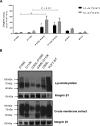
Impact of the introduction of the G305L mutation into the G112K and D109A scaffolds. A, [3H]MTX influx was assessed over 1 min at pH 5.5 and 37 °C at concentrations of 0.5 and 50 μm. Data are represented as percentage ± S.E. (error bars) of PCFT-WT from three independent experiments. A one-way ANOVA compared D109A or G112K with their respective G305L double mutants. *, p < 0.05. An analysis also compared G112K/G305L with the G112K single mutant at the low concentration indicated as a; p < 0.01. B, in this Western blotting, PCFT expression at the cell surface is indicated in the top row, and expression in the crude membrane extract is indicated in the bottom row, each with integrin β1 as the loading control. The blot is representative of three independent experiments.
Figure 8.
Figure 8.
Impact of the introduction of the G305L mutation into the P314C-D109A scaffold and the accessibility of the P314C residue to MTSEA-biotin. A, [3H]MTX influx was assessed at 37 °C and pH 5.5 over 1 min at concentrations of 0.5 and 50 μm. Data are represented as percentage ± S.E. of PCFT-WT influx from three independent experiments. A one-way ANOVA compared the D109A or D109A/P314C mutants with the triple mutant, D109A/P314C/G305L. ****, p < 0.0001. B, in this Western blotting, the top panel indicates the MTSEA-biotinylated protein, the middle panel shows the Lys-biotinylated protein assessed with EZ-Link Sulfo-NHS-LC, and the bottom panel is PCFT in the crude membrane extract. Integrin β1 serves a loading control for both the Lys-biotinylated and crude membrane proteins. The images are representative of three independent experiments.
Figure 9.
Figure 9.
A homology model of PCFT depicting the locations of the Asp-109, Gly-305, and Ser-196 residues. A, a planar view of PCFT. B, a view into the protein from the extracellular compartment. The Asp-109 residue is indicated in blue, the Gly-305 residue in green, and the Ser-196 residue in yellow. C, [3H]MTX influx was assessed at 37 °C over 1 min at pH 5.5 at concentrations of 0.5 and 50 μm in cells harboring the D109A mutation alone or in combination with the S196L or S196F mutation. Data are percentage ± S.E. (error bars) of PCFT-WT from three independent experiments. A one-way ANOVA compared D109A with the corresponding double mutants. **, p < 0.01. D, PCFT expression at the cell surface assessed with EZ-Link Sulfo-NHS-SS-Biotin is indicated in the top panel, and expression in the crude membrane extract is indicated in the bottom panel, each with integrin β1 as the loading control. The images are representative of three independent experiments.

References

    1. Zhao R., Aluri S., and Goldman I. D. (2017) The proton-coupled folate transporter (PCFT-SLC46A1) and the syndrome of systemic and cerebral folate deficiency of infancy: Hereditary folate malabsorption. Mol. Aspects Med. 53, 57–72 10.1016/j.mam.2016.09.002 - DOI - PMC - PubMed
    1. Qiu A., Jansen M., Sakaris A., Min S. H., Chattopadhyay S., Tsai E., Sandoval C., Zhao R., Akabas M. H., and Goldman I. D. (2006) Identification of an intestinal folate transporter and the molecular basis for hereditary folate malabsorption. Cell 127, 917–928 10.1016/j.cell.2006.09.041 - DOI - PubMed
    1. Visentin M., Diop-Bove N., Zhao R., and Goldman I. D. (2014) The intestinal absorption of folates. Annu. Rev. Physiol. 76, 251–274 10.1146/annurev-physiol-020911-153251 - DOI - PMC - PubMed
    1. Zhao R., Qiu A., Tsai E., Jansen M., Akabas M. H., and Goldman I. D. (2008) The proton-coupled folate transporter (PCFT): impact on pemetrexed transport and on antifolate activities as compared to the reduced folate carrier. Mol. Pharmacol. 74, 854–862 10.1124/mol.108.045443 - DOI - PMC - PubMed
    1. Matherly L. H., Hou Z., and Gangjee A. (2018) The promise and challenges of exploiting the proton-coupled folate transporter for selective therapeutic targeting of cancer. Cancer Chemother. Pharmacol. 81, 1–15 10.1007/s00280-017-3473-8 - DOI - PMC - PubMed

Publication types