. 2019 Mar 11;9(1):4131.
doi: 10.1038/s41598-019-40425-9.

Capsaicin Protects Against Cisplatin Ototoxicity by Changing the STAT3/STAT1 Ratio and Activating Cannabinoid (CB2) Receptors in the Cochlea

Affiliations

Capsaicin Protects Against Cisplatin Ototoxicity by Changing the STAT3/STAT1 Ratio and Activating Cannabinoid (CB2) Receptors in the Cochlea

Puspanjali Bhatta et al. Sci Rep. .

Abstract

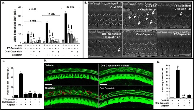
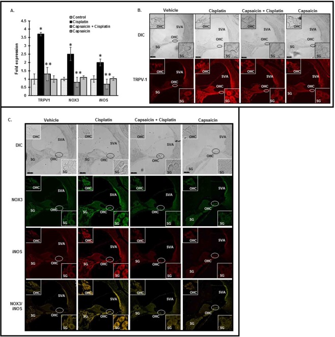
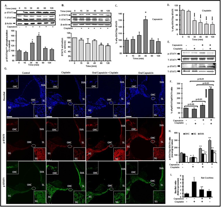
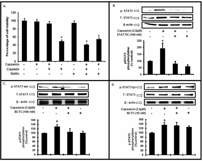
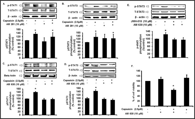
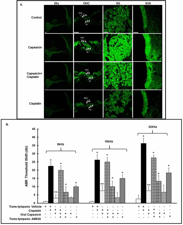
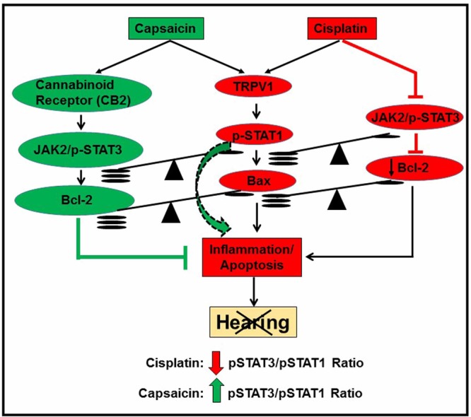
Capsaicin, the spicy component of hot chili peppers activates the TRPV1 pain receptors, and causes rapid desensitization. Capsaicin also ameliorates cisplatin-induced nephrotoxicity. Cisplatin, a commonly used anti-neoplastic agent for solid tumors causes significant hearing loss, nephrotoxicity and peripheral neuropathy. Upregulation of cochlear TRPV1 expression is related to cisplatin-mediated ototoxicity. Here we report that direct TRPV1 activation by localized trans-tympanic (TT) or oral administration of capsaicin (TRPV1 agonist) prevents cisplatin ototoxicity by sustained increased activation of pro-survival transcription factor signal transducer and activator of transcription (STAT3) in the Wistar rat. Cisplatin treatment produced prolonged activation of pro-apoptotic Ser727 p-STAT1 and suppressed Tyr705-p-STAT3 for up to 72 h in the rat cochlea. Our data indicate that capsaicin causes a transient STAT1 activation via TRPV1 activation, responsible for the previously reported temporary threshold shift. Additionally, we found that capsaicin increased cannabinoid receptor (CB2) in the cochlea, which leads to pro-survival Tyr705-p-STAT3 activation. This tilts the delicate balance of p-STAT3/p-STAT1 towards survival. Furthermore, capsaicin mediated protection is lost when CB2 antagonist AM630 is administered prior to capsaicin treatment. In conclusion, capsaicin otoprotection appears to be mediated by activation of CB2 receptors in the cochlea which are coupled to both STAT1 and STAT3 activation.


Conflict of interest statement

The authors declare no competing interests.

Figures

Figure 1
Figure 1
Capsaicin inhibits cisplatin-induced hearing loss. (A) We recorded ABR threshold shifts in naïve Wistar rats, pre-treated with either trans-tympanic capsaicin (0.1 µM, 50 µl) or TT-PBS (50 µl), oral PBS or oral capsaicin (20 mg/kg), 24 h prior to cisplatin (11 mg/kg, i.p). We measured post treatment ABR thresholds at 72 h after cisplatin and they showed significant increase with cisplatin alone, which capsaicin attenuated at all frequencies tested. Black arrows indicate significant decrease in threshold shifts when compared to cisplatin. (*Indicates significant difference from control; **indicates significant difference from cisplatin treatment, p < 0.05, n = 9). (B) SEM studies performed on rat cochleae show disruption of the stereociliary bundles indicating damage to the OHCs by cisplatin which was significantly ameliorated by pre-treatment with capsaicin. (C) Quantitative analysis of outer hair cell damage in the basal turn in the SEM images. (*Indicates significant difference from control; **indicates significant difference from cisplatin treatment, p < 0.05, n = 4). (D) Cochlear whole mount preparations from animals stained with myosin VIIA and imaged by confocal microscopy indicate that cisplatin caused hair cell loss in the basal turn (indicated by red arrows), while capsaicin pre-treatment was protective. Scale bar is 25 µm. (E) Quantitative analysis of presence or absence of outer hair cells from the basal turn of the whole mount. (*Indicates significant difference from control; **indicates significant difference from cisplatin treatment, p < 0.05, n = 4).
Figure 2
Figure 2
Capsaicin inhibits cisplatin-induced stress and inflammation in the cochlea. We pretreated male Wistar rats with trans-tympanic capsaicin (0.1 µM, 50 µl) 24 h prior to cisplatin (11 mg/kg). We collected -the cochleae 72 h post cisplatin, and saved these specimens either in RNA later or fixed with freshly prepared 4% paraformaldehyde, decalcified and processed them for mid-modiolar sections. (A) Quantitative q-PCR analyses from total cochlear RNA indicate that cisplatin treatment increased the relative mRNA expression of TRPV1, NOX3 and iNOS, that was inhibited by trans-tympanic pre-treatment with capsaicin. (B,C) We used mid-modiolar sections for immunofluorescent staining of TRPV1 (B), NOX3 and iNOS (C). Cisplatin increased TRPV1, NOX3 and iNOS staining in the OC, SL, SV and SG, which with trans-tympanic capsaicin pretreatment blocked. Images shown are 20X and the insets were imaged at 100x by confocal microscopy. Scale bar is 100 µm. Data presented in (A) represent the mean ± S.E.M. of cochleae from four animals from each group. Asterisk (*) indicates statistically significant difference from vehicle, while (**) indicates significant difference from the cisplatin group (p < 0.05).
Figure 3
Figure 3
Capsaicin activates both STAT1 and STAT3, while cisplatin activates STAT1 and suppresses pro-survival STAT3. (A,B) UB/OC1 cells treated with capsaicin (2.5 µM) demonstrated significantly increased Tyr705 p-STAT3 after 30 and 45 min (A). Cisplatin (2.5 µM) treatment suppressed the Tyr705–p-STAT3 (B) in a sustained and significant manner at 120 minutes. (C) Capsaicin treatment significantly increased the ratio of p-STAT3/p-STAT1 in a transient manner at 45 minutes. (D) Cisplatin treatment persistently and significantly decreased the p-STAT3/p-STAT1 ratio. (E,F) UB/OC1 cells were pretreated with capsaicin (2.5 µM) for 30 minutes followed by cisplatin (2.5 µM) treatment for 45 minutes. Cisplatin increases p-STAT1 significantly, with no change in p-STAT3 phosphorylation, while capsaicin increased p-STAT1 and p-STAT3 significantly. Capsaicin + cisplatin treatment decreased p-STAT1 significantly with a concomitant significant increase in p-STAT3 activation. Graphical analyses of p-STAT3/p-STAT1 ratio indicates that cisplatin significantly decreased the p-STAT3/p-STAT1 ratio compared to control, capsaicin significantly increased p-STAT3/p-STAT1 ratio compared to cisplatin. Capsaicin + cisplatin group showed a significant increase in p-STAT3/p-STAT1 ratio compared to all the groups, possibly due to significant increase in p-STAT3 activation and significant decrease in p-STAT1 activation. (G) Mid-modiolar sections of cochleae from rats treated with sterile PBS, i.p. (vehicle), capsaicin (20 mg/kg) or cisplatin (11 mg/Kg, i.p) for 72 hours were immunolabeled for Ser727-p-STAT1 (green) or Tyr705-p-STAT3 (red) and imaged using confocal microscopy (insets: 100X magnification). Cisplatin increased Ser727-p-STAT1 staining at 72 h post administration, while capsaicin treatment did not alter p-STAT1 compared to control in the cochlea. Capsaicin (72 h) increased Tyr705-p-STAT3 staining, while cisplatin (72 h) suppressed the Tyr705-p-STAT3 staining. Asterisks (*) indicate statistically significant difference from 0 min or control cells (p < 0.05); double asterisks (**) indicate statistically significant difference from cisplatin (p < 0.05); (n = 4). Scale bar is 100 µm. (H) Graphical representation of p-STAT3/p-STAT1 ratio of the different cochlear regions as analyzed by image J software. (I) Cochlear gene expression by q-PCR indicates that cisplatin treatment increased the Bax/Bcl-2 ratio, which was abrogated by capsaicin pre-treatment.
Figure 4
Figure 4
STAT3 is essential for capsaicin mediated protection and is TRPV1 independent. (A) We pretreated UB/OC-1 cells with a selective inhibitor of STAT3 (STATTIC, 100 nM) for 45 min, followed by capsaicin (2.5 µM) for 45 min and then cisplatin (20 µM) for 48 h. We assessed the percentage of cell viability by MTS assay. Treatment of STATTIC (100 nM) alone did not induce cell death whereas treatment with cisplatin induced significant cell death. Capsaicin (2.5 µM) protected against cisplatin (20 µM) induced cell death. However, pretreatment with STATTIC (100 nM) (i.e after the inhibition of STAT3), capsaicin could not protect against cisplatin-induced cell death. Asterisk (*) indicate statistically significant difference from control (p < 0.05, n = 8), (**) indicates statistically significant difference from cisplatin (p < 0.05, n = 8). (B) We pretreated UB/OC-1 cells with STATTIC (100 nM) for 45 min, followed by capsaicin (2.5 µM) for 45 min. Pretreatment of STATTIC inhibited the phosphorylation of Tyr705 p-STAT3. Asterisk (*) indicate significant difference from control (p < 0.05, n = 4). (C,D) We pretreated UB/OC-1 cells with BCTC (100 nM) for 45 min followed by capsaicin (2.5 µM) for 45 min. Western blots show that inhibition of TRPV1 reduced the Ser727 p-STAT1 (C) but not Tyr705 p-STAT3 (D). Western blotting: we loaded all samples serially as depicted on the gel. The grouping of blots, reflects the same blot re-probed with different antibodies. β-actin is used as loading control. None of the blots have lanes taken from different parts of the blot. Thick borders denote separate antibody probes for the same gel.
Figure 5
Figure 5
Cannabinoid Receptor CB2 activation is essential for capsaicin induced otoprotection. (A,B) To determine the role of CB1 receptor in capsaicin induced cell survival, we pretreated UB/OC-1 cells with CB1 antagonist, AM281 (10 µM) for 30 min followed by capsaicin (2.5 µM) for 45 min. Western blots showed that inhibition of CB1 receptors (by AM281) did not affect either the capsaicin-mediated Ser727 p-STAT1 (A) or Tyr705 p-STAT3 (B). Asterisk (*) indicates statistically significant change from control. (p < 0.001, n = 4). (C,D) To determine the role of CB2 receptor in capsaicin induced cell survival, we pretreated UB/OC-1 cells with CB2 antagonist, AM630 (10 µM) for 30 min followed by the capsaicin (2.5 µM) for 45 min. Western blots showed that inhibition of CB 2 receptors blocked the capsaicin-mediated Ser727 p-STAT1 (C) and Tyr705 p-STAT3 (D). Asterisk (*) indicates statistically significant change from control. (p < 0.001, n = 4). (E) We pretreated UB/OC-1 cells with or without CB2 antagonist, AM630 (10 µM) for 30 min followed by CB2 agonist, JWH-015 (10 µM) for 30 min. Western blots showed CB2 agonist (JWH-015) increased Tyr705 p-STAT3, and that inhibition of CB2 receptors (by AM630) reduced the JWH-015 -mediated Tyr705 p-STAT3 phosphorylation. Thus, this implicates CB2R in the protective signaling of STAT3. (Asterisk (*) indicates statistically significant change from control (p < 0.05, n = 4). (F) We pretreated UB/OC-1 cells with AM630 for 45 min, followed by capsaicin (2.5 µM) for 24 h. We assessed cell viability using MTS assay. Significant cell death occurred when cells were treated with CB2 antagonist, AM630 and capsaicin, implicating CB2 receptors in cell survival. Asterisk (*) indicate statistically significant change from control (p < 0.05, n = 4). Western blotting: we loaded all samples serially as depicted on the gel. The grouping of blots reflects the same blot re-probed with different antibodies. β-actin is used as loading control. None of the blots have lanes taken from different parts of the blot. Thick borders denote separate antibody probes for the same gel.
Figure 6
Figure 6
Cannabinoid receptors are expressed in the rat cochlea and CB2 receptor is integral to capsaicin mediated rescue of cisplatin-induced hearing loss. We treated male Wistar rats with oral PBS (1 ml) or oral capsaicin (10 mg/kg) and collected the cochleae 72 h later. (A) We immunolabeled mid-modiolar sections of rat cochleae for CB2R and imaged using confocal microscopy at 20X and 100X. We observed CB2R expression in outer hair cells (OHCs), supporting cells (SC’s), spiral ganglion cells (SG) and in the stria vascularis (SVA). Scale bar is 25 µm. (B) We pretreated rats were pretreated with AM630 (0.1 µM, TT), CB2R antagonist for 1 hour, then capsaicin (20 mg/kg, 1 ml) by oral gavage 24 hours prior to cisplatin administration (11 mg/kg i.p.). After 72 hours, we determined ABR threshold shifts at 8, 16 and 32 kHz. Pretreatment with capsaicin (20 mg/Kg solution) by oral gavage significantly reduced the threshold shifts caused by cisplatin. CB2 receptor antagonist AM630 (TT) abrogated the capsaicin protection against cisplatin. AM630 (TT) alone also caused ABR threshold shifts. Asterisks (*) and (**) indicate statistically significant increases from PBS and from cisplatin-treated animals, respectively (p < 0.05, n = 4).
Figure 7
Figure 7
Oral capsaicin pre-treatment does not affect cisplatin’s chemotherapeutic ability in the SCID mouse xenograft model. We injected twenty seven SCID mice with UMSCC-10b cells (1 × 106 cells) subcutaneously and divided them into four treatment groups: Oral PBS + PBS (i.p, 1 ml) (n = 8), cisplatin (2 mg/kg, i.p) (n = 7), oral capsaicin (0.5 mg/kg) + cisplatin (2 mg/kg, i.p) (n = 7) and oral capsaicin alone (n = 5). We administered drugs by oral gavage and/or i.p injections every other day (three times a week, once the tumors were palpable, until the end of the study. We monitored tumor growth closely. Oral capsaicin + Cisplatin and the cisplatin treated mice showed suppressed tumor size (CF and I) and weight (J). Capsaicin treatment alone decreased tumor size and weight, but not significantly. We show representative images of the tumor bearing mice and excised tumors at the end of the study from each group in (AH). Line graphs and histograms in (I,J) represent mean ± SEM from each treatment group. Asterisk (*) indicates statistically significant difference (p < 0.05) from vehicle-treated mice.
Figure 8
Figure 8
A summary of capsaicin protection against cisplatin ototoxicity. This chart summarizes the proposed molecular mechanisms underlying capsaicin mediated protection against cisplatin-induced ototoxicity. It appears that otoprotection mediated by capsaicin is produced by activation of CB2 receptors which further activates STAT3. Activation of STAT1 by capsaicin contributes to the transient inflammatory response without recruitment of apoptotic pathways as previously observed in vivo (as shown by the green dotted arrow). The transient inflammation through the activation of STAT1 desensitizes the TRPV1 receptors, decreasing the presence of STAT1 to be activated by cisplatin. Such phenomenon can mitigate the undesired inflammation further initiated by cisplatin. The net protective action of capsaicin could result from an increase in the JAK2 and STAT3/STAT1 ratio in cells in the cochlea, abrogating the negative impact of cisplatin on this ratio.

References

    1. Suresh D, Srinivasan K. Tissue distribution & elimination of capsaicin, piperine & curcumin following oral intake in rats. Indian J Med Res. 2010;131:682–691. - PubMed
    1. Han D, et al. Activation of cannabinoid receptor type II by AM1241 protects adipose-derived mesenchymal stem cells from oxidative damage and enhances their therapeutic efficacy in myocardial infarction mice via Stat3 activation. Oncotarget. 2017;8:64853–64866. doi: 10.18632/oncotarget.17614. - DOI - PMC - PubMed
    1. Barsotti A, et al. MK-954 (losartan potassium) exerts endothelial protective effects against reperfusion injury: evidence of an e-NOS mRNA overexpression after global ischemia. Atherosclerosis. 2001;155:53–59. doi: 10.1016/S0021-9150(00)00533-5. - DOI - PubMed
    1. Caterina MJ, Julius D. The vanilloid receptor: a molecular gateway to the pain pathway. Annu Rev Neurosci. 2001;24:487–517. doi: 10.1146/annurev.neuro.24.1.487. - DOI - PubMed
    1. Holzer P. Capsaicin as a tool for studying sensory neuron functions. Adv Exp Med Biol. 1991;298:3–16. doi: 10.1007/978-1-4899-0744-8_1. - DOI - PubMed

Publication types

MeSH terms