. 2019 Mar 11;9(1):4026.
doi: 10.1038/s41598-019-40436-6.

Hyaluronic acid inhibition by 4-methylumbelliferone reduces the expression of cancer stem cells markers during hepatocarcinogenesis

Affiliations

Hyaluronic acid inhibition by 4-methylumbelliferone reduces the expression of cancer stem cells markers during hepatocarcinogenesis

Caecilia H C Sukowati et al. Sci Rep. .

Abstract

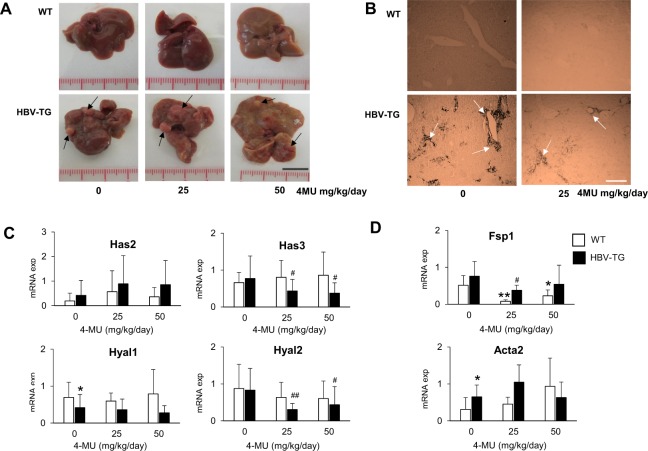
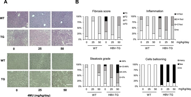
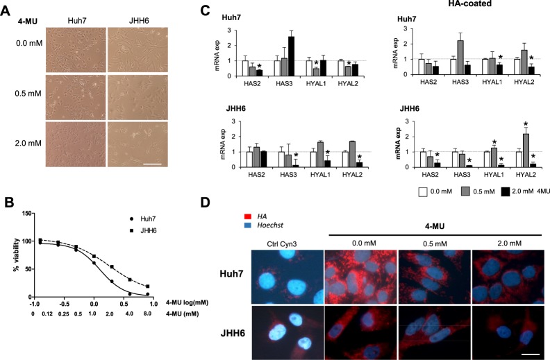
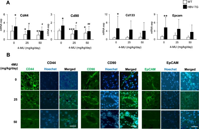
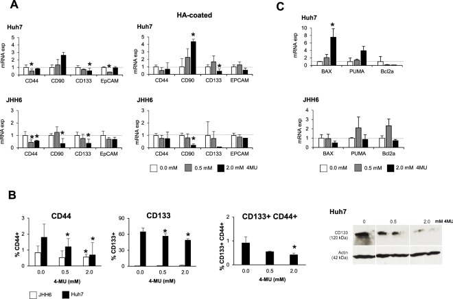
Hyaluronic acid (HA) is a glycosaminoglycan of extracellular matrix related to cell surface which interacts with various cell types. To understand the role of HA during hepatocarcinogenesis, we assessed the effect of the inhibition of HA deposition and its association with heterogeneous hepatocellular carcinoma (HCC) cells. In this study, we used transgenic mice C57BL/6J-Tg(Alb1HBV)44Bri/J (HBV-TG) and normal C57BL/6 J (WT) for in vivo study, while HCC cells Huh7 and JHH6 as in vitro models. Both models were treated with an HA inhibitor 4-methylumbelliferone (4MU). We observed that 4MU treatments in animal model down-regulated the mRNA expressions of HA-related genes Has3 and Hyal2 only in HBV-TG but not in normal WT. As observed in vivo, in HCC cell lines, the HAS2 mRNA expression was down-regulated in Huh7 while HAS3 in JHH6, both with or without the presence of extrinsic HA. Interestingly, in both models, the expressions of various cancer stem cells (CD44, CD90, CD133, and EpCAM) were also decreased. Further, histological analysis showed that 4MU treatment with dose 25 mg/kg/day reduced fibrosis, inflammation, and steatosis in vivo, in addition to be pro-apoptotic. We concluded that the inhibition of HA reduced the expressions of HA-related genes and stem cells markers in both models, indicating a possible modulation of cells-to-cells and cells-to-matrix interaction.


Conflict of interest statement

The authors declare no competing interests.

Figures

Figure 1
Figure 1
Effect of 4MU treatment in HBV-TG mice. (A) Macroscopic appearance of liver after 12 weeks treatment of 25 and 50 mg/kg/day 4MU (scale bar = 1 cm). (B) HA staining of the liver before and after treatment. Magnification: 200X (scale bar = 300 µm). (C) Relative mRNA expressions of hyaluronan synthases Has2 and Has3, and hyaluronidases Hyal1 and Hyal2. (D) Relative mRNA expressions of fibrosis and ECM genes Fsp1 and Acta2. Each mRNA expression of target genes was normalized to reference genes Gapdh and Actb. Statistical analysis: *p < 0.05, **p < 0.01 compared to WT 0 mg/kg/day, #p < 0.05, ##p < 0.01 compared to TG 0 mg/kg/day. WT = wild type mice, HBV-TG = HBV-transgenic mice.
Figure 2
Figure 2
4MU treatment improves histology in HBV-TG mice. (A) Representative pictures of H&E staining (upper panel) and reticulum staining (lower panel) (scale bar = 200 µm). (B) The percentage of fibrosis score, steatosis grade, and inflammation scores in the liver. WT = wild type mice, HBV-TG = transgenic mice.
Figure 3
Figure 3
Effect of 4MU treatment in HCC cells JHH6 and Huh7. (A) Morphological cellular changes after treatment (scale bar = 200 µm). (B) Dose-dependent cytotoxicity of 4MU with concentration ranging from 0.0–8.0 mM by MTT test. Concentration was shown as log[mM]. (C) Relative mRNA expressions of hyaluronan synthases HAS2 and HAS3, and hyaluronidases HYAL1 and HYAL2 after treatment of 0.5 and 2.0 mM of 4MU. mRNA expression was normalized to reference genes 18S and ACTB. Statistical analysis: *p < 0.05, **p < 0.01 compared to untreated control of each cell line (0.0 mM = 1.0) (D) Presence of cytoplasmic HA (scale bar = 20 µm).
Figure 4
Figure 4
4MU treatment reduces the expressions of CSC markers in mouse model. (A) Relative mRNA expressions of CSC markers Cd44, Cd90, Cd133, and Epcam in HBV-TG and WT mice. Statistical analysis: *p < 0.05, **p < 0.01 compared to WT 0 mg/kg/day, #p < 0.05, ##p < 0.01 compared to TG 0 mg/kg/day. (B) Representative of immunofluorescence images of protein CD44, CD90, and EpCAM in HBV-TG (scale bar = 50 µm). WT = wild type mice, HBV-TG = HBV-transgenic mice.
Figure 5
Figure 5
4MU treatment reduces the expressions of CSC markers in HCC cells JHH6 and Huh7. (A) Relative mRNA expressions of CSC markers CD44, CD90, CD133, and EpCAM after treatment. (B) Flow cytometric analysis on the number of CD44+, CD133+, and CD133+CD44+after treatment (left). Western blot representative of CD133 protein of Huh7 (right). Full-length Western blot is presented in Supplementary Fig. S1. (C) Relative mRNA expressions of apoptosis genes after treatment. mRNA expression was normalized to reference genes 18S and ACTB. Statistical analysis: *p < 0.05 vs. untreated control of each cell line (0.0 mM = 1.0).

References

    1. Bray F, Ren JS, Masuyer E, Ferlay J. Global estimates of cancer prevalence for 27 sites in the adult population in 2008. Int. J. Cancer. 2013;132:1133–1145. doi: 10.1002/ijc.27711. - DOI - PubMed
    1. Llovet JM, Burroughs A, Bruix J. Hepatocellular carcinoma. Lancet. 2003;362:1907–1917. doi: 10.1016/S0140-6736(03)14964-1. - DOI - PubMed
    1. Toole BP. Hyaluronan: from extracellular glue to pericellular cue. Nat. Rev. Cancer. 2004;4:528–539. doi: 10.1038/nrc1391. - DOI - PubMed
    1. Toole BP. Hyaluronan promotes the malignant phenotype. Glycobiology. 2002;12:37R–42R. doi: 10.1093/glycob/12.3.37R. - DOI - PubMed
    1. Alaniz L, Garcia M, Rizzo M, Piccioni F, Mazzolini G. Altered hyaluronan biosynthesis and cancer progression: an immunological perspective. Mini Rev. Med. Chem. 2009;9:1538–1546. doi: 10.2174/138955709790361485. - DOI - PubMed

Publication types