. 2019 Apr;29(4):313-329.
doi: 10.1038/s41422-019-0153-8. Epub 2019 Mar 11.

Vitamin B12 modulates Parkinson's disease LRRK2 kinase activity through allosteric regulation and confers neuroprotection

Affiliations

Vitamin B12 modulates Parkinson's disease LRRK2 kinase activity through allosteric regulation and confers neuroprotection

Adam Schaffner et al. Cell Res. 2019 Apr.

Abstract

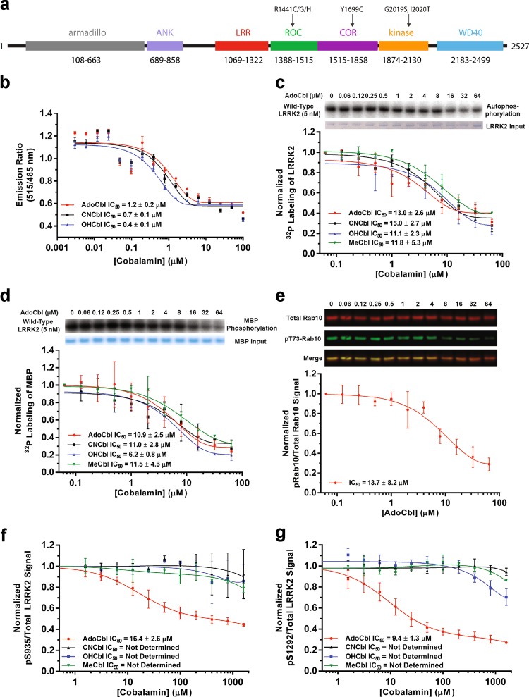
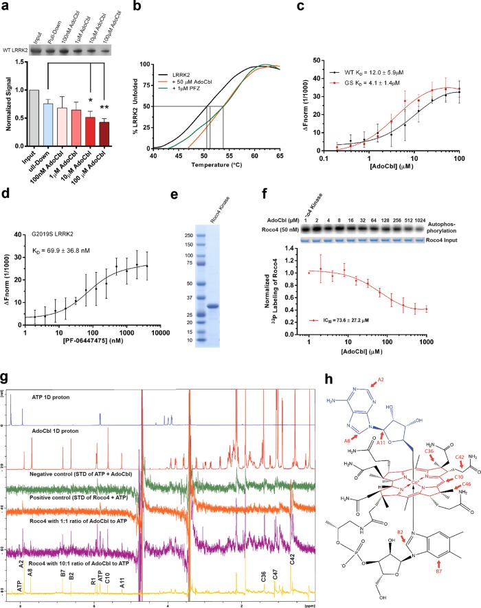
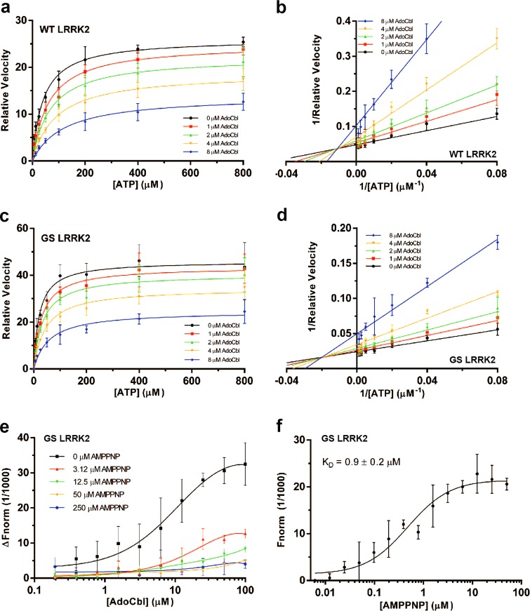
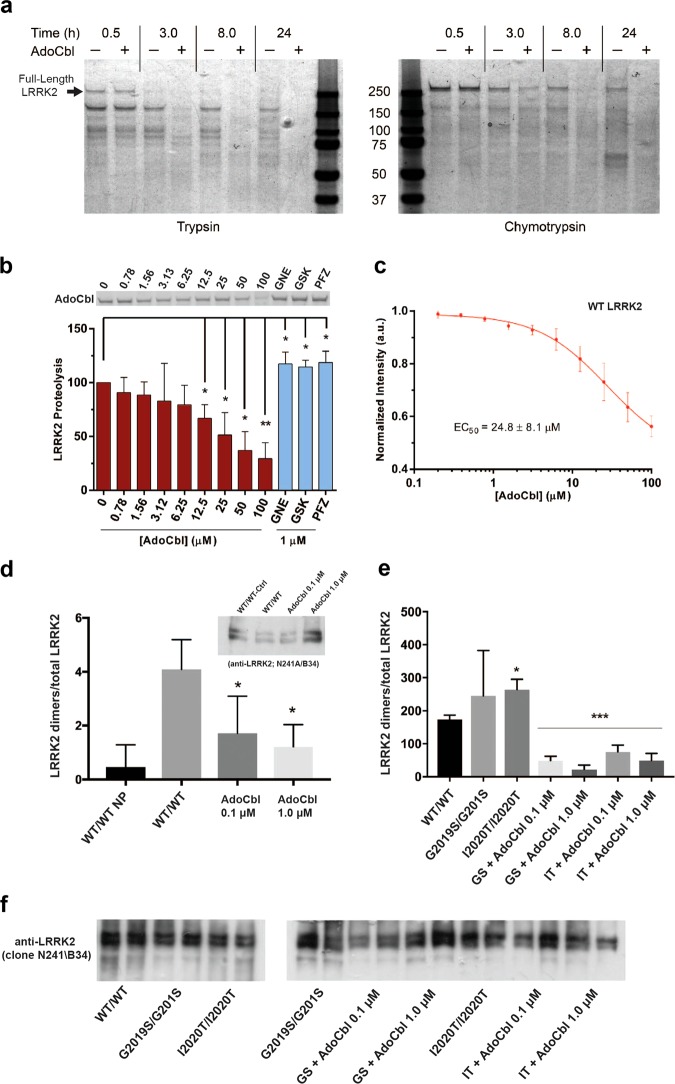
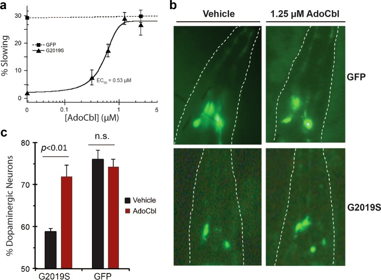
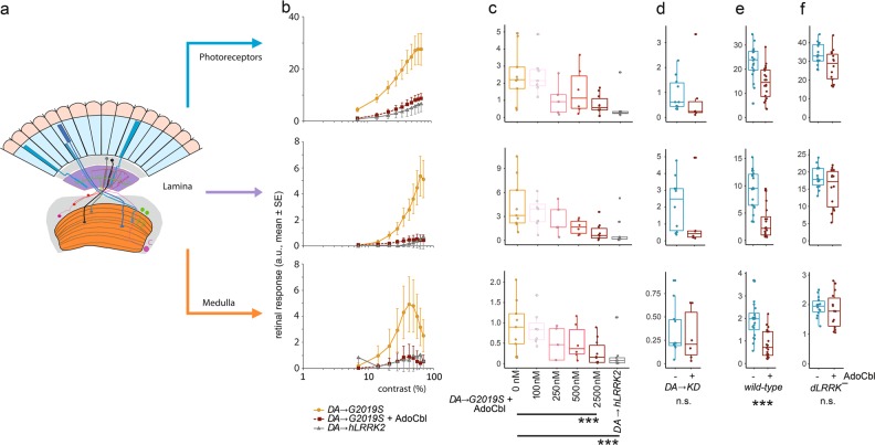
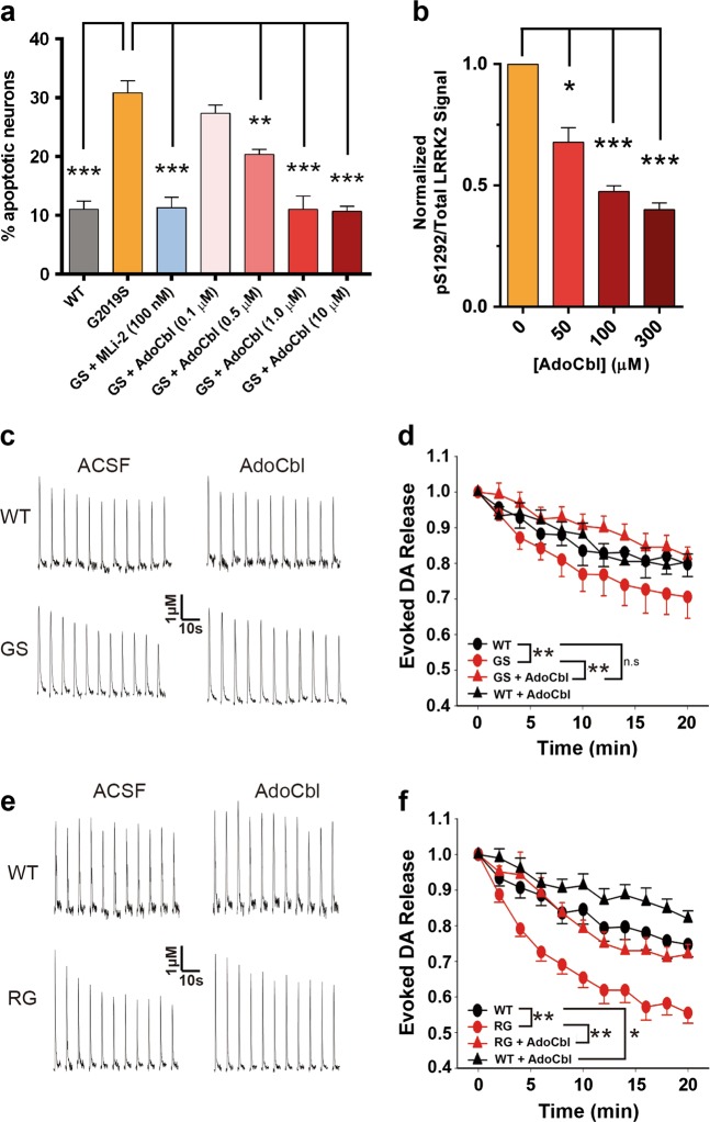
Missense mutations in Leucine-Rich Repeat Kinase 2 (LRRK2) cause the majority of familial and some sporadic forms of Parkinson's disease (PD). The hyperactivity of LRRK2 kinase induced by the pathogenic mutations underlies neurotoxicity, promoting the development of LRRK2 kinase inhibitors as therapeutics. Many potent and specific small-molecule LRRK2 inhibitors have been reported with promise. However, nearly all inhibitors are ATP competitive-some with unwanted side effects and unclear clinical outcome-alternative types of LRRK2 inhibitors are lacking. Herein we identify 5'-deoxyadenosylcobalamin (AdoCbl), a physiological form of the essential micronutrient vitamin B12 as a mixed-type allosteric inhibitor of LRRK2 kinase activity. Multiple assays show that AdoCbl directly binds LRRK2, leading to the alterations of protein conformation and ATP binding in LRRK2. STD-NMR analysis of a LRRK2 homologous kinase reveals the contact sites in AdoCbl that interface with the kinase domain. Furthermore, we provide evidence that AdoCbl modulates LRRK2 activity through disrupting LRRK2 dimerization. Treatment with AdoCbl inhibits LRRK2 kinase activity in cultured cells and brain tissue, and prevents neurotoxicity in cultured primary rodent neurons as well as in transgenic C. elegans and D. melanogaster expressing LRRK2 disease variants. Finally, AdoCbl alleviates deficits in dopamine release sustainability caused by LRRK2 disease variants in mouse models. Our study uncovers vitamin B12 as a novel class of LRRK2 kinase modulator with a distinct mechanism, which can be harnessed to develop new LRRK2-based PD therapeutics in the future.


Conflict of interest statement

The authors declared no competing interest.

Figures

Fig. 1
Fig. 1
AdoCbl inhibits LRRK2 kinase activity. a Domain structure of LRRK2. b Dose-response curves of brain-purified flag-tagged LRRK2 kinase as a function of different forms of cobalamin. Phosphorylation is quantified by measuring TR-FRET emission ratios of fluorescein-LRRKtide and a Terbidium-labeled pLRRKtide antibody. c Dose-response curves of strep-tagged LRRK2 autophosphorylation or d phosphorylation of myelin basic protein as a function of different forms of cobalamin. e Dose-response curve of strep-tagged LRRK2-G2019S phosphorylation of purified Rab10 as a function of AdoCbl. f Dose-response curves of pS935/Total LRRK2 and g pS1292/Total LRRK2 after treatment with different forms of cobalamin in MEF cells derived from LRRK2-G2019S BAC transgenic mice. Data from each replicate were normalized to LRRK2 phosphorylation without cobalamin treatment. All data points represent the mean (±s.d.) of three biological replicates
Fig. 2
Fig. 2
Direct binding of AdoCbl to LRRK2 protein. a Binding of strep-tagged LRRK2 to AdoCbl-agarose in the presence of AdoCbl. Input represents the amount of protein that was added to beads, while pull-down denotes the amount of protein left on the beads after washes. Significance was calculated by one-way ANOVA using the mean (±s.d.) of three biological replicates. *p ≤ 0.05, **p ≤ 0.005. b Thermal shift assays showing melting temperatures of strep-LRRK2 in the presence of AdoCbl or PF-06447475. c Microscale thermophoretic analysis of the interaction between AdoCbl or d PF-06447475 with strep-tagged LRRK2. e Coomassie stained SDS-PAGE of the Roco4 kinase domain purified from E. coli. f Dose-response curve of Roco4 kinase activity as a function of AdoCbl. g ATP STD-NMR shows direct binding of AdoCbl to the Roco4 kinase domain and competition with ATP. From top to bottom, the spectra are as follows: 1D 1 H NMR of ATP (blue), AdoCbl (red), STD negative control with ATP + AdoCbl only (green), STD positive control with ATP and Roco4 kinase domain (orange), STD of AdoCbl and Roco4 kinase domain with 1:1 ratio of AdoCbl to ATP (purple), and STD of AdoCbl and Roco4 kinase domain with 10:1 ratio of AdoCbl to ATP (yellow). AdoCbl protons showing strong STD signals are labeled with assignment. All experiments were collected at 4 °C on a Bruker 800 MHz spectrometer equipped with a cryoprobe. h Protons with strong STD signals (highlighted in red) mapped onto the structure of AdoCbl. The NMR assignment and nomenclature of vitamin B12 is from Summers et al. Data points in a, c, d, f represent the mean (±s.d.) of three biological replicates
Fig. 3
Fig. 3
AdoCbl exhibits a mixed-mode of inhibition. a Michaelis-Menten kinetics curves of full-length Invitrogen flag-tagged LRRK2-WT and c LRRK2-G2019S as a function of AdoCbl. Relative velocity represents the value of pS1292/Total LRRK2 after 20 min of reaction time at 30 °C (during the linear reaction rate), as detected by western blot and quantified by densitometry. b Lineweaver-Burk plots of LRRK2-WT and d LRRK2-G2019S kinetics data. e Microscale thermophoretic analysis of the interaction between AdoCbl and strep-tagged LRRK2 in the presence of increasing concentrations of AMP-PNP. Fluorescently-labeled strep-tagged LRRK2 was pre-incubated with AMP-PNP before binding between LRRK2 and AdoCbl was measured by MST. f Microscale thermophoretic analysis of the interaction between AMP-PNP and LRRK2, showing a KD of 0.9 μM. Data points represent the mean (±s.d.) of three biological replicates
Fig. 4
Fig. 4
AdoCbl causes a LRRK2 conformational change and destabilizes LRRK2 dimers. a Coomassie stained SDS-PAGE showing limited proteolysis analysis using a 10:1 molar ratio of LRRK2: Trypsin (left panel) and LRRK2: Chymotrypsin (right panel). Proteolysis was performed at 30 °C with or without 50 µM AdoCbl and reactions were quenched at the indicated times by the addition of sample loading buffer. The observed data were consistent across three biological replicates. b Limited proteolysis of LRRK2-WT by trypsin in the presence of increasing concentrations of AdoCbl, or 1 µM LRRK2 kinase inhibitor. Proteolysis was performed for 90 min at 30 °C. Shown is a representative SDS-PAGE of full-length LRRK2, in which bands were quantified and values were normalized to LRRK2 proteolysis without AdoCbl. c The peak intrinsic fluorescence of LRRK2 (339 nm) was measured as a function of AdoCbl. Strep-tagged LRRK2 was incubated with indicated concentrations of AdoCbl for 30 min prior to fluorescence measurements. Significance was measured by one-way ANOVA. *p ≤ 0.05, **p ≤ 0.005. d HEK293T cells co-expressing BirA-WT (biotin ligase) and AP-WT LRRK2 (acceptor peptide) were lysed following a biotin pulse to label dimeric LRRK2, and extracts bound to streptavidin-coated ELISA plates. LRRK2 was detected using anti-LRRK2 conjugated to HRP (clone N241A/B34) and expressed as a ratio of total LRRK2 levels detected by ELISA in parallel plates coated with total LRRK2 antibodies (clone c41-2). In the plot, “WT/WT NP” refers to cells expressing WT LRRK2 dimers that were harvested without receiving a biotin pulse (“no pulse”). AdoCbl significantly reduced levels of dimeric WT-LRRK2. Sub-panel shows representative immunoblot of parallel extracts detected with anti-LRRK2 (clone N241A/B34). BirA-LRRK2 represents the top band, and AP-LRRK2 the bottom band. e HEK293T cells expressing BirA- G2019S or I2020T mutant LRRK2 together with AP-G2019S or I2020T LRRK2, and dimeric LRRK2 quantified by ELISA. Treatment with AdoCbl significantly reduces dimeric mutant LRRK2. *p < 0.05 compared to WT/WT-LRRK2; ***p < 0.001 compared to G2019S-LRRK2 dimers or I2020T-LRRK2 dimers alone. f Representative immunoblot of parallel extracts detected with anti-LRRK2 (clone N241A/B34). BirA-LRRK2 represents the top band, and AP-LRRK2 the bottom band
Fig. 5
Fig. 5
AdoCbl rescues mutant human LRRK2-induced behavioral defects and dopaminergic neurodegeneration in C. elegans. a AdoCbl dose dependently rescues the loss of basal slowing response in transgenic hLRRK2-G2019S C. elegans. Age-synchronized nematodes expressing GFP marker only or additionally hLRRK2-G2019S in dopaminergic neurons were treated with either vehicle or AdoCbl in liquid culture during the larval stage L1 to L4 (3 days), followed by growth on NGM plates for 3 days prior to behavior assay. Basal slowing response was assayed on NGM plates using an unbiased machine-vision analysis system (WormLab) as the percent slowing in body bends per 20 s in the presence vs. the absence of bacterial lawn. Data represent the mean (±s.d.) of three biological replicates, each with 20–25 worms per treatment condition. b AdoCbl treatment attenuated the loss of dopaminergic neurons induced by hLRRK2-G2019S in C. elegans. Representative fluorescence images of dopaminergic neurons (CEP neurons within the outlined head region) in transgenic C. elegans expressing GFP marker only or additionally hLRRK2-G2019S following treatment with either vehicle or 1.25 µM AdoCbl. Age-synchronized nematodes were treated with either vehicle or AdoCbl in liquid culture during the larval stage L1 to L4 (3 days), followed by growth on NGM plates for 9 days. GFP-tagged dopaminergic neurons in live animals were counted under a fluorescence microscope. c Quantification of percent dopaminergic neurons survived. Data are presented as the mean (±s.d.) of three biological replicates, each with approximately 30–50 worms per treatment condition. P< 0.01, Student’s t-test. n.s., not statistically significant
Fig. 6
Fig. 6
AdoCbl rescues deficits in Drosophila visual physiology induced by the dopaminergic expression of human LRRK2-G2019S. a Outline of the retinal neural network of Drosophila, with three main neuronal layers: photoreceptors, lamina neurons, and medulla neurons (Modified after Afsari et al.) b Contrast response functions (CRFs) for the photoreceptors, lamina neurons and medulla neurons show that the dopaminergic expression of hLRRK2-G2019S (DAG2019S) flies have a much stronger response than either the DAhLRRK2 or the DAG2019S which have been fed 2.5 μM AdoCbl. c Dose-response curve for the effect of AdoCbl on the DAG2019S flies, shows a 50% reduction in phenotypes by 250–500 nM AdoCbl, with almost complete rescue by 2.5 μM AdoCbl. d There is no effect of 2.5 μM AdoCbl on flies with dopaminergic expression of kinase-dead hLRRK2-G2019S-K1906M (DAKD). e The visual response of flies with wild-type dLRRK2 is reduced by 2.5 μM AdoCbl. f Applying 2.5 μM AdoCbl to dLRRK¯ transheterozygote flies (in which the Drosophila LRRK2 homolog has been knocked out) has no statistically significant effect. Data represent the mean (±s.e.) and the dots represent the number of flies tested. In c, statistical analysis from Tukey Post-hoc tests on the first principal component of a PCA, which accounted for 88% of the variance (Supplementary information, Fig. S9). df analysis by MANOVA. n.s. not significant; ***p < 0.001). Boxes correspond to the median ± quartiles. Dots indicate data from individual flies. dLRRK¯ genotype: dLRRKe03680/dLRRKex1; wild-type genotype: wa/w1118
Fig. 7
Fig. 7
AdoCbl prevents LRRK2-G2019S induced neurotoxicity and rescues deficits in dopamine transmission in LRRK2-PD mouse models. a Quantification of percent apoptotic neurons after LRRK2 overexpression and treatment with MLi-2 or AdoCbl. Cortical neurons were co-transfected with LRRK2 and a GFP reporter. Transfected neurons displaying apoptotic nuclear morphology were counted 48 h after transfection using DAPI. Apoptotic neurons were defined as those having condensed fragmented chromatin comprised of two or more apoptotic bodies. Data represent the mean (±s.d.) from n = 3 biological replicates of triplicate coverslips. Significance was measured by one-way ANOVA. b Quantification of pS1292/Total LRRK2 after brain slice tissue from LRRK2-G2019S BAC-transgenic mice were treated with AdoCbl. One mouse brain provided enough slices to test each treatment condition one time. Three mouse brains were used in total, resulting in three biological replicates. Data are the mean (±s.d.) and significance was measured by one-way ANOVA c Voltammetric traces of striatal DA release evoked at 2-min intervals from G2019S, or e R1441G, and WT controls after 2 h treatment with control vehicle (water), or with 300 µM AdoCbl. d Summary of DA release sustainability for G2019S mice (n = 9 sites) or f R1441G (n = 10 sites) compared to WT controls. Data are expressed as the mean (±SEM) and were analyzed by two-way ANOVA with Bonferroni’s post hoc analysis. For all figures, *p ≤ 0.01, **p ≤ 0.001, ***p ≤ 0.0001

Comment in

References

    1. Dickson DW, et al. Neuropathology of non-motor features of Parkinson disease. Park. & Relat. Disord. 2009;15(Suppl. 3):S1–S5. - PubMed
    1. Lees AJ, Hardy J, Revesz T. Parkinson’s disease. Lancet. 2009;373:2055–2066. - PubMed
    1. Funayama M, et al. A new locus for Parkinson’s disease (PARK8) maps to chromosome 12p11.2-q13.1. Ann. Neurol. 2002;51:296–301. - PubMed
    1. Paisán-Ruíz C, et al. Cloning of the gene containing mutations that cause PARK8-linked Parkinson’s disease. Neuron. 2004;44:595–600. - PubMed
    1. Zimprich A, et al. Mutations in LRRK2 cause autosomal-dominant parkinsonism with pleomorphic pathology. Neuron. 2004;44:601–607. - PubMed

Publication types