Review
. 2019 Jun;15(6):346-366.
doi: 10.1038/s41581-019-0129-4.

Glycosylation in health and disease

Affiliations
Review

Glycosylation in health and disease

Colin Reily et al. Nat Rev Nephrol. 2019 Jun.

Erratum in

Abstract

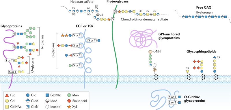
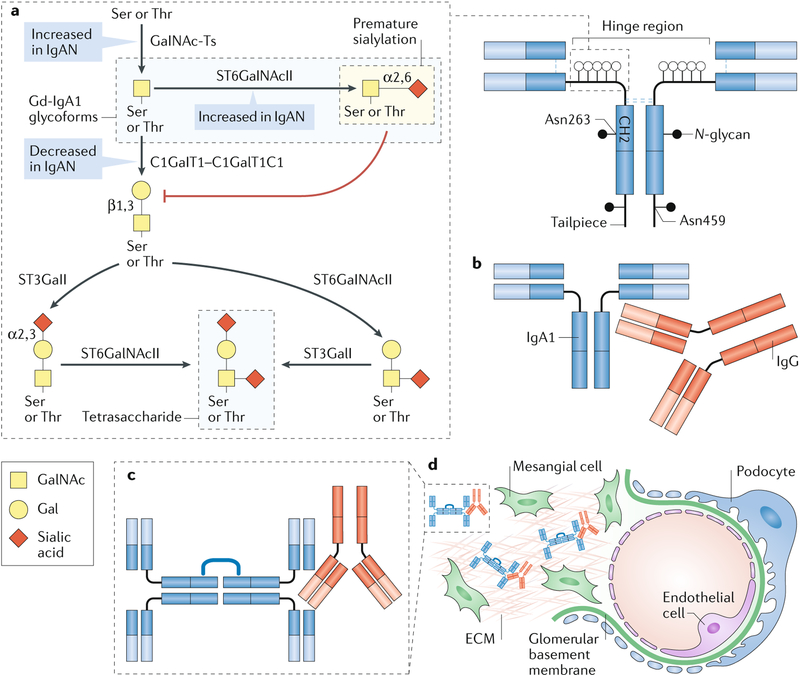
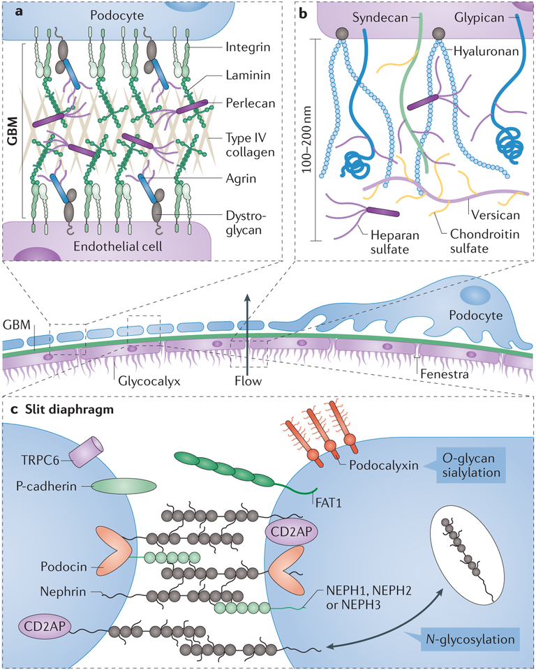
The glycome describes the complete repertoire of glycoconjugates composed of carbohydrate chains, or glycans, that are covalently linked to lipid or protein molecules. Glycoconjugates are formed through a process called glycosylation and can differ in their glycan sequences, the connections between them and their length. Glycoconjugate synthesis is a dynamic process that depends on the local milieu of enzymes, sugar precursors and organelle structures as well as the cell types involved and cellular signals. Studies of rare genetic disorders that affect glycosylation first highlighted the biological importance of the glycome, and technological advances have improved our understanding of its heterogeneity and complexity. Researchers can now routinely assess how the secreted and cell-surface glycomes reflect overall cellular status in health and disease. In fact, changes in glycosylation can modulate inflammatory responses, enable viral immune escape, promote cancer cell metastasis or regulate apoptosis; the composition of the glycome also affects kidney function in health and disease. New insights into the structure and function of the glycome can now be applied to therapy development and could improve our ability to fine-tune immunological responses and inflammation, optimize the performance of therapeutic antibodies and boost immune responses to cancer. These examples illustrate the potential of the emerging field of 'glycomedicine'.


Conflict of interest statement

Competing interests

J.N. and M.B.R. are co-founders of Reliant Glycosciences, LLC. The other authors declare no competing interests.

Figures

Fig. 1 |
Fig. 1 |. Major types of glycosylation in humans.
Glycans can be covalently attached to proteins and lipids to form glycoconjugates; glycans in these compounds are classified according to the linkage to the lipid, glycan or protein moieties. Glycoproteins consist of glycans and glycan chains linked to nitrogen and oxygen atoms of amino acid residues and are thus termed N-glycans and O-glycans, respectively. N-glycans consist of N-acetylglucosamine (GlcNAc) attached by a β1-glycosidic linkage to the nitrogen atom of the amino group of Asn (N) at the consensus glycosylation motif Asn-X-Ser/Thr (in which X denotes any amino acid except for Pro). These branched and highly heterogeneous N-glycan structures consist of a core glycan containing two GlcNAc residues and three mannose (Man) residues. Perhaps the most diverse form of protein glycosylation is O-glycosylation, in which glycans attach to the oxygen atom of the hydroxyl groups of Ser (S) or Thr (T) residues. O-glycans can be further subclassified on the basis of the initial sugar attached to the protein and the additional sugar structures added to the initial glycan. For example, mucin-type O-glycosylation denotes that the initial glycan is N-acetylgalactosamine (GalNAc); mucin-type glycans can be further classified on the basis of the glycans attached to the initial GalNAc. Other types of O-glycans, such as O-linked fucose (Fuc) and O-linked Man, often occur in specific proteins or protein domains, such as epidermal growth factor (EGF) repeats, thrombospondin type I repeats (TSR) or dystroglycan. N-glycans and O-glycans are often capped with negatively charged sialic acid. O-GlcNAc is a unique type of O-glycosylation that is synthesized by O-GlcNAc transferase; it occurs in the cytosol and nucleus. Proteoglycans represent a major class of glycoproteins that are defined by long glycosaminoglycan (GAG) chains attached to proteins through a tetrasaccharide core consisting of glucuronic acid (GlcA)–galactose (Gal)–Gal–xylose (Xyl); this carbohydrate core is attached to the hydroxyl group of Ser at Ser-Gly-X-Gly amino acid motifs. Proteoglycan GAGs can be further classified according to the number, composition and degree of sulfation of their repeating disaccharide units; common GAGs include heparan sulfate, chondroitin sulfate and dermatan sulfate. Glycosylphosphatidylinositol (GPI)-anchored glycoproteins represent another major class of glycoconjugates. These glycoproteins are linked at the carboxyl terminus through a phosphodiester linkage to phosphoethanolamine attached to a trimannosyl-nonacetylated glucosamine (Man3-GlcN) core; the GlcN residue is linked to phosphatidylinositol, which is embedded in the cell membrane. Glycosphingolipids are a class of glycoconjugate in which glycans, such as Gal or glucose (Glc), are attached to cellular membrane lipids. Another major class of glycans is represented by GAGs that are not attached to protein cores, such as hyaluronan, which is synthesized at the plasma membrane by sequential addition of GlcA and GlcNAc. IdoA, iduronic acid. Adapted with permission from ref., Springer Nature Limited and from ref., Stanley, P. Golgi glycosylation. Cold Spring Harb. Perspect. Biol. 0033, a005199 (2005), with permission from Cold Spring Harbor Laboratory Press.
Fig. 2 |
Fig. 2 |. N-glycan biosynthesis in the secretory pathway.
N-glycan synthesis is initiated in the endoplasmic reticulum (ER) by the en bloc transfer of a lipid-glycan precursor (that is, glucose (Glc)3 mannose (Man)9 N-acetylglucosamine (GlcNAc)2 bound to dolichol phosphate) to Asp by the multisubunit oligosaccharyltransferase (OST). The glucose residues are sequentially removed by two α-glucosidases (α-Glc I–II) and an initial Man residue is removed by the ER α-mannosidase (ER α-Man). After a quality-control checkpoint, the glycoprotein moves to the Golgi apparatus for additional trimming by α-mannosidase I and II (α-Man I–II) and further glycan modifications. A cis-to-trans distribution of glycosidases and transferases — GlcNAc-transferase I–IV (GnT-I–IV), β1,4 galactosyltransferases (Gal-T), α2,3 sialyltransferase (α2,3, Sialyl-T) and α2,6 sialyltransferase (α2,6 Sialyl-T) — facilitates further processing by these carbohydrate-modifying enzymes to create a plethora of N-glycoforms that often terminate with sialic acid moieties. The final site-specific N-glycan composition is affected by the expression levels of glycosyltransferases, the accessibility of the glycoprotein glycosylation sites and the length of time during which the glycoprotein remains in the ER and Golgi apparatus. Gal, galactose.
Fig. 3 |
Fig. 3 |. Functional impact of variable Igg Fc glycan composition.
a | Immunoglobulin G (IgG) has two heavy and two light chains, and its crystallizable fragment (Fc) region can bind to Fcγ receptors and some proteins of the complement system. The IgG Fc region contains two N-glycans, one per heavy chain, attached at Asn297; these glycans contribute to the structural integrity of the Fc region and to its interactions with Fc receptors and complement. The Fc glycans in the IgG molecule are biantennary glycans with variable content of fucose (Fuc), bisecting N-acetylglucosamine (GlcNAc), galactose (Gal) and sialic acid; most IgG molecules are fucosylated. The glycan composition of IgG affects its biological activity; for example, Gal-deficient IgG glycoforms, which have been associated with chronic inflammatory diseases, can activate the lectin complement pathway. b | IgG glycoforms with Gal-deficient glycans are pro-inflammatory. c | IgG glycoforms with sialylated glycans are considered to be anti-inflammatory. Man, mannose.
Fig. 4 |
Fig. 4 |. Structure and glycosylation of human IgA.
Human immunoglobulin A (IgA) occurs in two subclasses, IgA1 and IgA2. a | The amino acid sequence is very similar for both subclasses, but the IgA1 heavy chain contains additional amino acids in the hinge region. Each heavy chain of IgA1 also contains two N-glycans, one in the CH2 domain (Asn263) and one in the tailpiece (Asn459). Although human IgA2 does not contain O-glycans, it can have more N-glycans per heavy chain than IgA1. b | The IgA1 hinge region is composed of two octapeptide repeats, which include nine Ser and Thr residues that are potential O-glycosylation sites; usually 3–6 of these residues are O-glycosylated and the most common IgA1 glycoforms have 4–5 O-glycans in the hinge region. c | The O-glycan composition of normal circulating human IgA1 is variable but usually consists of a core 1 disaccharide structure with N-acetylgalactosamine (GalNAc) in β1,3-linkage with galactose (Gal); each of these monosaccharides can be sialylated. d | The CH2 site of N-glycosylation contains digalactosylated biantennary glycans with or without a bisecting N-acetylglucosamine (GlcNAc), but it is not usually fucosylated. e | By contrast, the tailpiece N-glycosylation site contains fucosylated glycans. Fuc, fucose; Man, mannose.
Fig. 5 |
Fig. 5 |. ST6galI and abnormal sialylation in cancer.
N-glycans with terminal α2,6-sialylation are synthesized by the β-galactoside α−2,6-sialyltransferase 1 (ST6GalI). Both upregulation of ST6GalI and increase in α2,6-sialylation are observed in many types of cancers and are associated with negative patient outcomes. In fact, α2,6-sialylation is involved in the regulation of many key proteins that are known to contribute to cell survival and metastasis in cancer. a | Hypoxia increases the expression and activity of ST6GalI, leading to enhanced α2,6-sialylation; in turn, increased sialylation upregulates HIF1α and the expression of pro-survival HIF1α target genes, such as growth factors and glucose transporters. b | The death receptor FAS, also known as CD95, is a target of ST6GalI, and its sialylation inhibits the initiation of apoptotic signalling and subsequent receptor internalization. Increased expression of ST6GalI prevents FAS ligand (FASL)-induced apoptosis through FAS. c | Increased expression of ST6GalI in cancer cell lines enhances the α2,6-sialylation of epidermal growth factor receptor (EGFR), which increases its tyrosine kinase activity and the phosphorylation of its targets. α2,6-Sialylation enhances the activity of EGFR, both at baseline and after cell activation, and leads to increased activation of pro-growth and survival genes. Moreover, cells in which ST6GalI is overexpressed, leading to enhanced α2,6-sialylation, are protected against cell death induced by the anticancer drug gefitinib, an EGFR inhibitor. d | In cells with low ST6GalI expression and reduced α2,6 sialylation, prolonged activation of tumour necrosis factor (TNF) receptor 1 (TNFR1) by TNF leads to receptor internalization, caspase activation and cell death. This apoptotic cell death pathway is prevented by enhanced α2,6-sialylation of TNFR. e | α2,6-Sialylation of β1 integrin in the Golgi apparatus by ST6GalI results in hypersialylation, which inhibits β1 integrin binding to matrix proteins such as type I collagen and fibronectin and prevents downstream signalling. These signals maintain cell quiescence, and their disruption due to enhanced sialylation leads to increased cell motility and invasion, which promotes cancer cell metastasis.
Fig. 6 |
Fig. 6 |. Aberrant O-glycosylation in IgA nephropathy.
Immunoglobulin A (IgA) nephropathy (IgAN) is an autoimmune disease that is thought to result from a four-hit process. a | Hit 1: increased production of circulating galactose (Gal)-deficient IgA1 (Gd-IgA1), in which some O-glycans do not contain Gal. The first step in the glycosylation of the hinge region of IgA1 is the addition of N-acetylgalactosamine (GalNAc) to Ser or Thr residues by a GalNAc-transferase (GalNAc-T) to form the terminal GalNAc (Tn) antigen. Premature sialylation of the terminal GalNAc by α-GalNAc α−2, 6-sialyltransferase 2 (ST6GalNAcII) can block subsequent glycosylation; however, most often GalNAc is galactosylated by the glycosyltransferase GalNAc 3β-galactosyltransferase 1 (C1GalT1). The chaperone C1GalT1C1 is required for appropriate expression and function of C1GalT1. After galactosylation, GalNAc, Gal or both sugars may be sialylated; ST6GalNAcII adds sialic acid to GalNAc and β-galactoside α−2,3-sialyltransferase 1 (ST3GalI) sialylates Gal; the largest glycan of circulatory IgA1 is a tetrasaccharide. In many patients with IgAN, the number of O-glycosylated residues in the hinge region of IgGA1 is also increased82,83. Increased initiation of glycosylation by GalNAc-Ts, premature sialylation of GalNAc by ST6GalNAcII and decreased galactosylation by C1GalT1 might each contribute to the formation of Gd-IgA1, the key autoantigen in IgAN. b | Hit 2: production of IgG autoantibodies that are specific for Gd-IgA1. c | Hit 3: formation of circulating Gd-IgA1–IgG immune complexes. For simplicity, IgA1 is shown as a dimer; IgA1 monomers are the main circulating molecular form, but polymeric IgA1 is the predominant molecular form of Gd-IgA1 in the circulation. d | Hit 4: glomerular deposition of immune complexes. Other serum proteins, such as complement, are likely to be involved in the formation of the pathogenic immune complexes that are deposited in the glomeruli, activate resident mesangial cells and cause renal injury. ECM, extracellular matrix.
Fig. 7 |
Fig. 7 |. Glycoconjugates and glomerular filtration.
Glomerular filtration occurs in the glomerulus, and endothelial cells and podocytes are the principal cells involved in this process. Mesangial cells have a key role in maintaining and supporting the endothelial and podocyte glomerular filtration barrier, in part through the production of extracellular matrix proteins, and in regulating blood pressure. a | The glomerular basement membrane (GBM) is found at the interface between endothelial cells and podocytes; the GBM contains a network of type IV collagen, negatively charged heparan sulfate proteoglycans, laminins and several other extracellular matrix proteins. Loss of proteoglycans that contain heparan sulfate, such as agrin or perlecan, occurs in some glomerular diseases, including minimal change disease and membranous nephropathy. Glycosylation of membrane-associated α-dystroglycan has an important role in the interaction between the cells that line the GBM and the extracellular matrix through its interaction with laminin. b | The endothelial glycocalyx extends far from the plasma membrane of the glomerular endothelial cells, creating a physical barrier of glycoproteins, glycosaminoglycans and proteoglycans across the fenestrae. The glycan components of the glycocalyx have a key role in maintaining its mechanical and structural integrity and enabling its proper function as part of the glomerular filtration unit. Hyaluronan extends from the cell surface, whereas chondroitin sulfate and heparin sulfate are attached to extracellular matrix proteins, such as versican and perlecan, or membrane proteins such as syndecan and glypican. Together, these molecules make a dense negatively charged glycocalyx. c | The structure of the slit diaphragm, the key component of the glomerular molecular filter, relies heavily on the cell-surface adhesion protein nephrin; appropriate N-glycosylation of nephrin is critical for its surface expression and function. Sialylation of the cell-surface sialoglycoprotein podocalyxin also has a key role in podocyte morphogenesis and structural integrity. Many other cell-surface glycoproteins are involved in the formation of the slit diaphragm, including P-cadherin, podocin, nephrin-like proteins 1–3 (NEPH1–NEPH3), protocadherin fat 1 (FAT1) and transient receptor protein 6 (TRPC6). CD2-associated protein (CD2AP) is an adaptor molecule that can bind to the cytoplasmic domain of nephrin.

References

    1. Varki A Biological roles of glycans. Glycobiology 27, 3–49 (2017). - PMC - PubMed
    2. This paper provides a comprehensive overview of glycobiology and the various roles of glycans.

    1. Varki A et al. Symbol nomenclature for graphical representations of glycans. Glycobiology 25, 1323–1324 (2015). - PMC - PubMed
    1. Laine RA A calculation of all possible oligosaccharide isomers both branched and linear yields 1.05 × 10(12) structures for a reducing hexasaccharide: the Isomer Barrier to development of single-method saccharide sequencing or synthesis systems. Glycobiology 4, 759–767 (1994). - PubMed
    1. Spiro RG Protein glycosylation: nature, distribution, enzymatic formation, and disease implications of glycopeptide bonds. Glycobiology 12, 43R–56R (2002). - PubMed
    1. Gagneux P & Varki A Evolutionary considerations in relating oligosaccharide diversity to biological function. Glycobiology 9, 747–755 (1999). - PubMed

Publication types

MeSH terms