. 2019 Feb 27:41:5.
doi: 10.1186/s41021-019-0121-z. eCollection 2019.

Standard protocol for the total red blood cell Pig-a assay used in the interlaboratory trial organized by the Mammalian Mutagenicity Study Group of the Japanese Environmental Mutagen Society

Affiliations

Standard protocol for the total red blood cell Pig-a assay used in the interlaboratory trial organized by the Mammalian Mutagenicity Study Group of the Japanese Environmental Mutagen Society

Satsuki Chikura et al. Genes Environ. .

Abstract

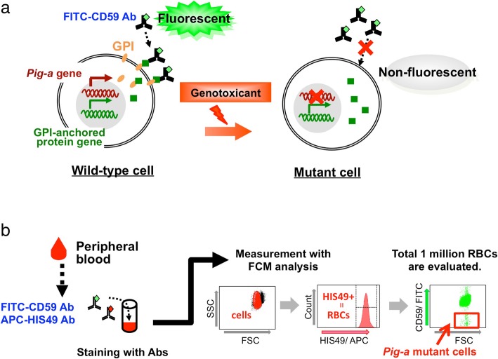
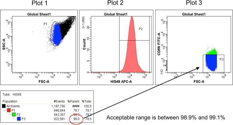
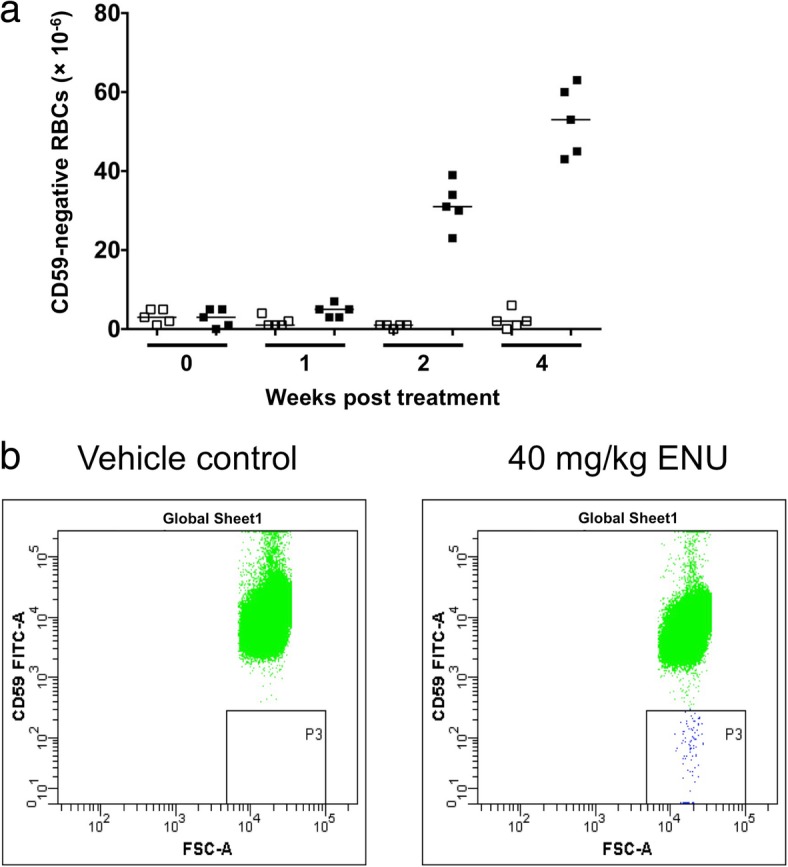
The Pig-a assay, a promising tool for evaluating in vivo genotoxicity, is based on flow cytometric enumeration of red blood cells (RBCs) that are deficient in glycosylphosphatidylinositol anchor protein. Various approaches for measuring Pig-a mutant cells have been developed, particularly focusing on measuring mutants in peripheral RBCs and reticulocytes (RETs). The Pig-a assay on concentrated RETs-the PIGRET assay-has the potential to detect genotoxicity in the early stages of a study. To verify the potential and usefulness of the PIGRET assay for short-term testing, we conducted an interlaboratory trial involving 16 laboratories organized by the Mammalian Mutagenicity Study Group of the Japanese Environmental Mutagen Society (MMS/JEMS). The collaborating laboratories assessed the mutagenicity of a total of 24 chemicals in rats using a single-treatment design and standard protocols for conducting the Pig-a assay on total RBCs (the RBC Pig-a assay) and the PIGRET assay. Here, we describe the standard protocol for the RBC Pig-a assay in detail.

Keywords: CD59; Flow cytometry; Glycosylphosphatidylinositol; HIS49; In vivo gene mutation; Pig-a assay; Red blood cells.


Conflict of interest statement

Not applicable.The authors declare that they have no competing interests.Springer Nature remains neutral with regard to jurisdictional claims in published maps and institutional affiliations.

Figures

Fig. 1
Fig. 1
Principle of the Pig-a assay and flow cytometry analysis. a Pig-a is an essential gene for synthesis of the glycosylphosphatidylinositol (GPI) anchor. In wild-type cells, GPI anchors and CD59, a GPI-anchored protein marker, are synthesized independently and GPI tethers CD59 to the cell surface. However, in Pig-a mutant cells, CD59 proteins on the cell surface are reduced because GPI anchors are not synthesized due to Pig-a gene mutation(s). Thus, Pig-a mutant cells do not react with FITC-conjugated anti-CD59 antibodies while wild-type cells react to the antibodies and fluoresce. b Peripheral blood is stained with fluorescent-labeled antibodies. Cells are gated by light scatter and then analyzed by flow cytometry for HIS49 rat erythroid marker expression. HIS49-positive cells are further analyzed for CD59 expression and Pig-a mutant cells are detected as the FITC-negative population
Fig. 2
Fig. 2
Plots for the RBC Pig-a assay. Create dot plots of FSC vs SSC (Plot 1) and FSC vs FITC (Plot 3) and a histogram of APC fluorescence (Plot 2). Set FSC and SSC in the log scale. When using digital flow cytometry systems, set area scaling factors to appropriate values and use the area measurement
Fig. 3
Fig. 3
Creating gates and adjustments with the non-stained control sample. Create P1, P2, and P3 gates and confirm that the P2 and P3 gates are subpopulations of the P1 and P2 gates, respectively. Display only the parent’s population on the subsequent plot (e.g., only the P1 population is displayed in Plot 2)
Fig. 4
Fig. 4
Gate adjustment with the CD59 single-stain sample. Adjust the P2 gate to exclude the negative cell population; insure that %parent of P2 is below 0.5%
Fig. 5
Fig. 5
Gate adjustment with the HIS49 single-stain sample. Adjust the height of the P3 gate to include 99.0 ± 0.1% of the CD59-negative cell population. Make sure that the P3 gate reaches the x-axis. If applicable, select “Bi-exponential” for the y-axis (FITC intensity scaling) in the Plot Inspector
Fig. 6
Fig. 6
Example RBC Pig-a assay results and typical flow cytometer plots. a RBC Pig-a assays were conducted pre-treatment (0), and at 1, 2, and 4 weeks after a single administration of PBS (vehicle control, open symbols) or 40 mg/kg ENU (closed symbols). This figure was reproduced from Genes and Environment 36, 199–202, 2014 [20] and modified. b Typical plots measuring Pig-a mutant cells (CD59-negative cells) from vehicle control and ENU treatment groups

References

    1. Gollapudi BB, Lynch AM, Heflich RH, Dertinger SD, Dobrovolsky VN, Froetschl R, et al. The in vivo Pig-a assay: a report of the international workshop on genotoxicity testing (IWGT) workgroup. Mutat Res - Genet Toxicol Environ Mutagen. 2015;783:23–35. doi: 10.1016/j.mrgentox.2014.09.007. - DOI - PubMed
    1. Assessment and Control of DNA Reactive (Mutagenic) Impurities in Pharmaceuticals To Limit Potential Carcinogenic Risk Guidance for Industry, ICH M7(R1). 2018.
    1. Kawagoe K, Takeda J, Endo Y, Kinoshita T. Molecular cloning of murine Pig-a, a gene for GPI-anchor biosynthesis, and demonstration of interspecies conservation of its structure, function, and genetic locus. Genomics Academic Press. 1994;23:566–574. doi: 10.1006/geno.1994.1544. - DOI - PubMed
    1. Watanabe R, Inoue N, Westfall B, Taron CH, Orlean P, Takeda J, et al. The first step of glycosylphosphatidylinositol biosynthesis is mediated by a complex of PIG-A, PIG-H, PIG-C and GPI1. EMBO J European Molecular Biology Organization. 1998;17:877–885. doi: 10.1093/emboj/17.4.877. - DOI - PMC - PubMed
    1. Nishimura J, Murakami Y, Kinoshita T. Paroxysmal nocturnal hemoglobinuria: an acquired genetic disease. Am J Hematol. 1999;62:175–182. doi: 10.1002/(SICI)1096-8652(199911)62:3<175::AID-AJH7>3.0.CO;2-8. - DOI - PubMed

LinkOut - more resources