Review
. 2019 Feb 26:11:191-204.
doi: 10.1016/j.dadm.2019.01.002. eCollection 2019 Dec.

Harmonizing brain magnetic resonance imaging methods for vascular contributions to neurodegeneration

Affiliations
Review

Harmonizing brain magnetic resonance imaging methods for vascular contributions to neurodegeneration

Eric E Smith et al. Alzheimers Dement (Amst). .

Abstract

Introduction: Many consequences of cerebrovascular disease are identifiable by magnetic resonance imaging (MRI), but variation in methods limits multicenter studies and pooling of data. The European Union Joint Program on Neurodegenerative Diseases (EU JPND) funded the HARmoNizing Brain Imaging MEthodS for VaScular Contributions to Neurodegeneration (HARNESS) initiative, with a focus on cerebral small vessel disease.

Methods: Surveys, teleconferences, and an in-person workshop were used to identify gaps in knowledge and to develop tools for harmonizing imaging and analysis.

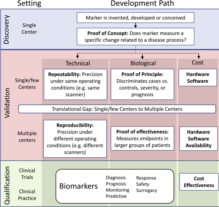
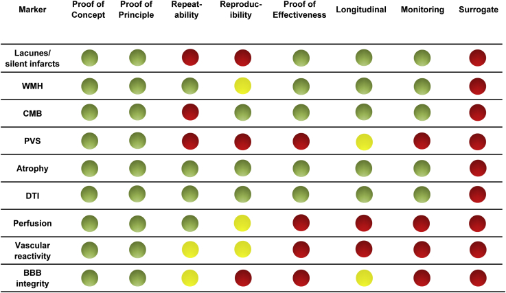
Results: A framework for neuroimaging biomarker development was developed based on validating repeatability and reproducibility, biological principles, and feasibility of implementation. The status of current MRI biomarkers was reviewed. A website was created at www.harness-neuroimaging.org with acquisition protocols, a software database, rating scales and case report forms, and a deidentified MRI repository.

Conclusions: The HARNESS initiative provides resources to reduce variability in measurement in MRI studies of cerebral small vessel disease.

Keywords: Cerebrovascular disease; Dementia; Magnetic resonance imaging; Radiology; Stroke.


Figures

Fig. 1
Fig. 1
Imaging biomarker development framework for cerebral small vessel disease.
Fig. 2
Fig. 2
Schematic overview of the neuroimaging biomarker development status for cerebral small vessel disease. The green light indicates validation data from two or more studies from independent research groups; yellow light indicates support from a single study or conflicting evidence from multiple studies; and red light indicates there is currently insufficient evidence. Abbreviations: WMH, white matter hyperintensities of presumed vascular origin; CMB, cerebral microbleeds; PVS, perivascular spaces; DTI, diffusion tensor imaging; BBB, blood-brain barrier. Proof of concept: evidence that the marker measures a specific change or process related to cerebral small vessel disease. Proof of principle/mechanism: evidence that the marker differs between patients with and without cerebral small vessel disease. Proof of effectiveness: evidence from larger scale multiple center studies that the marker differs between patients with and without cerebral small vessel disease. Repeatability: precision of repeated measurements under the same conditions using the same scanner. Reproducibility: replicate measurements on the same or similar objects (e.g., a phantom or human volunteers) in different locations using different scanners. Longitudinal: the rate of change over time has been defined. Monitoring: evidence that longitudinal changes in the marker are associated with progression of cerebral small vessel disease. Surrogate: evidence that changes in the marker are strongly associated with clinical outcomes in cerebral small vessel disease, such that changes in the marker could be considered a substitute for a clinical endpoint.

References

    1. Arvanitakis Z., Capuano A.W., Leurgans S.E., Bennett D.A., Schneider J.A. Relation of cerebral vessel disease to Alzheimer's disease dementia and cognitive function in elderly people: A cross-sectional study. Lancet Neurol. 2016;15:934–943. - PMC - PubMed
    1. Wardlaw J.M., Smith C., Dichgans M. Mechanisms of sporadic cerebral small vessel disease: Insights from neuroimaging. Lancet Neurol. 2013;12:483–497. - PMC - PubMed
    1. Vermeer S.E., Prins N.D., den Heijer T., Hofman A., Koudstaal P.J., Breteler M.M. Silent brain infarcts and the risk of dementia and cognitive decline. N Engl J Med. 2003;348:1215–1222. - PubMed
    1. Debette S., Beiser A., DeCarli C., Au R., Himali J.J., Kelly-Hayes M. Association of MRI markers of vascular brain injury with incident stroke, mild cognitive impairment, dementia, and mortality: the Framingham Offspring Study. Stroke. 2010;41:600–606. - PMC - PubMed
    1. Wardlaw J.M., Smith E.E., Biessels G.J., Cordonnier C., Fazekas F., Frayne R. Neuroimaging standards for research into small vessel disease and its contribution to ageing and neurodegeneration. The Lancet Neurol. 2013;12:822–838. - PMC - PubMed