. 2019 Jul;58(7):1134-1144.
doi: 10.1002/mc.22997. Epub 2019 Mar 11.

Tumor suppressor KIF1Bβ regulates mitochondrial apoptosis in collaboration with YME1L1

Affiliations

Tumor suppressor KIF1Bβ regulates mitochondrial apoptosis in collaboration with YME1L1

Koji Ando et al. Mol Carcinog. 2019 Jul.

Abstract

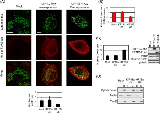
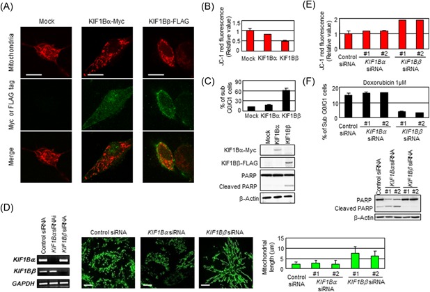
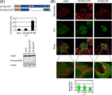
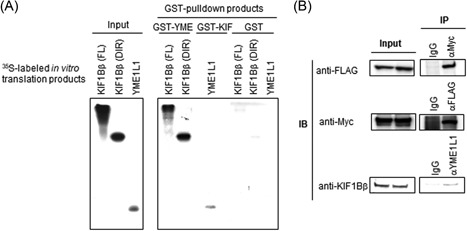
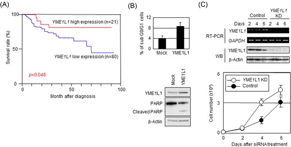
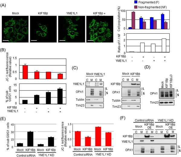
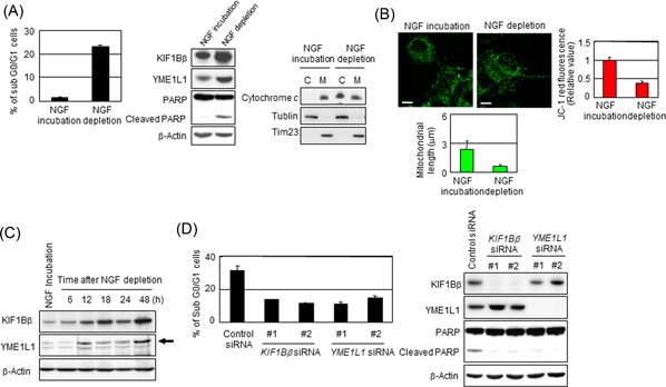
KIF1Bβ, a member of the kinesin superfamily of motor proteins, is a haploinsufficient tumor suppressor mapped to chromosome 1p36.2, which is frequently deleted in neural crest-derived tumors, including neuroblastoma and pheochromocytoma. While KIF1Bβ acts downstream of the nerve growth factor (NGF) pathway to induce apoptosis, further molecular functions of this gene product have largely been unexplored. In this study, we report that KIF1Bβ destabilizes the morphological structure of mitochondria, which is critical for cell survival and apoptosis. We identified YME1L1, a mitochondrial metalloprotease responsible for the cleavage of the mitochondrial GTPase OPA1, as a physical interacting partner of KIF1Bβ. KIF1Bβ interacted with YME1L1 through its death-inducing region, as initiated the protease activity of YME1L1 to cleave the long forms of OPA1, resulting in mitochondrial fragmentation. Overexpression of YME1L1 promoted apoptosis, while knockdown of YME1L1 promoted cell growth. High YME1L1 expression was significantly associated with a better prognosis in neuroblastoma. Furthermore, in NGF-deprived PC12 cells, KIF1Bβ and YME1L1 were upregulated, accompanied by mitochondrial fragmentation and apoptotic cell death. Small interfering RNA-mediated knockdown of either protein alone, however, remarkably inhibited the NGF depletion-induced apoptosis. Our findings indicate that tumor suppressor KIF1Bβ plays an important role in intrinsic mitochondria-mediated apoptosis through the regulation of structural and functional dynamics of mitochondria in collaboration with YME1L1. Dysfunction of the KIF1Bβ/YME1L1/OPA1 mechanism may be involved in malignant biological features of neural crest-derived tumors as well as the initiation and progression of neurodegenerative diseases.

Keywords: KIF1Bβ; YME1L1; mitochondrial fragmentation; neuroblastoma; tumor suppressor.


Conflict of interest statement

The authors declare that they have no conflict of interests.

Figures

Figure 1
Figure 1
Overexpression of KIF1Bβ induces mitochondrial fragmentation. A, Overexpression of KIF1Bβ results in mitochondrial fragmentation. HeLa cells were transfected with empty vector (Mock), KIF1Bα‐Myc, or KIF1Bβ‐FLAG expression vectors. After 48 hours, mitochondrial morphology was observed by indirect immunofluorescence using the BacMam‐GFP mitochondrial probe (green), Myc or FLAG tag antibodies (red). Scale bar, 25 µm. Average mitochondrial length is shown in a bar plot (n = 100). Data are presented as means ± SD (B) KIF1Bβ overexpression decreases membrane potential (ΔΨm). The average values of JC‐1 red fluorescence are shown as the normalized values by the value obtained from control. *P < 0.05 (n = 3). C, Overexpression of KIF1Bβ induces apoptosis. Cells in (A) were analyzed by flow cytometry (left panel) for the sub‐G0/G1 population. Cleavage of PARP was detected by Western blot analysis experiments using whole cell lysates (right panel). D, KIF1Bβ overexpression induces cytochrome c release from mitochondria to cytoplasm. Cells in (A) were fractionated into cytoplasmic (C) and mitochondrial (M) fractions and was subjected to immunoblotting. PARP, poly ADP‐ribose polymerase [Color figure can be viewed at wileyonlinelibrary.com]
Figure 2
Figure 2
Deficiency in KIF1Bβ yields mitochondrial fusion. A, Add‐back of KIF1Bβ reverts spontaneous mitochondrial fusion in NB‐1 cells. NB‐1 cells were transfected with empty vector (Mock), KIF1Bα‐Myc, or KIF1Bβ‐FLAG expression vectors. After 48 hours, mitochondria are visualized by MitoTrackerRed CMXRos. B, Add‐back of KIF1Bβ decreases membrane potential. The average values of JC‐1 red fluorescence are measured in NB‐1 cells and shown as the normalized values by the value obtained from control. *P < 0.05 (n = 3). C, Add‐back of KIF1Bβ attenuates drug resistance. NB‐1 cells transfected with KIF1Bα or KIF1Bβ were treated with doxorubicin. Apoptosis was analyzed by flow cytometry and immunoblotting. D, Knockdown of KIF1Bβ results in mitochondrial fusion. HeLa cells were transfected with a scramble siRNA (control) or two kinds of specific siRNAs against KIF1Bα or KIF1Bβ for 48 hours. Knockdown efficiency was confirmed by semiquantitative RT‐PCR (left panel). Morphological changes in mitochondria were observed by indirect immunofluorescence using the BacMam‐GFP mitochondrial probe (green). Scale bar, 25 µm. The average mitochondrial length was measured (right panel, n = 100). E, Knockdown of KIF1Bβ increases membrane potential ΔΨm. The values of JC‐1 red fluorescence in the cells in (D) were measured and shown as the normalized values by the value obtained from control. F, Knockdown of KIF1Bβ inhibits apoptosis induced by doxorubicin. After the siRNA knockdown, HeLa cells were treated with 1 µM doxorubicin for 48 hours. Apoptosis was analyzed by flow cytometry and immunoblotting. RT‐PCR, real‐time polymerase chain reaction; siRNA, small interfering RNA [Color figure can be viewed at wileyonlinelibrary.com]
Figure 3
Figure 3
The death‐inducing region of KIF1Bβ is essential for mitochondrial fragmentation. A, The death‐inducing region is essential for apoptosis induction. Schematic representation of GFP‐tagged KIF1Bβ full‐length and its death‐inducing region deletion mutant KIF1BβΔ3 is shown (top panel). HeLa cells were transfected with EGFP (control), KIF1BβΔ3‐GFP, or KIF1Bβ‐GFP for 48 hours and then subjected to flow cytometry (lower left panel) and immunoblotting (lower right panel). B, The overexpression of KIF1BβΔ3 does not induce mitochondrial fragmentation. EGFP, KIF1BβΔ3‐GFP, or KIF1Bβ‐GFP (green) was overexpressed in HeLa cells. Mitochondria were visualized by the BacMam‐RFP mitochondrial probe (red). Scale bar, 25 µm. Average mitochondrial length is shown in a bar plot (n = 100). DIR, death‐inducing region; EGFP, enhanced green fluorescent protein; Motor, motor domain; PARP, poly ADP‐ribose polymerase [Color figure can be viewed at wileyonlinelibrary.com]
Figure 4
Figure 4
Mitochondrial metalloprotease YME1L1 interacts with KIF1Bβ. A, Physical interaction between the death‐inducing region of KIF1Bβ and YME1L1 in vitro. [35S] labeled KIF1Bβ death‐inducing region (DIR), full‐length KIF1Bβ, or YME1L1 was incubated with recombinant GST‐YME1L1, GST‐KIF1Bβ, or GST negative control. GST proteins were analyzed by Western blot analysis and autoradiography. B, Physical interaction between KIF1Bβ and YME1L1 in vivo. Lysates from HeLa cells cotransfected with expression plasmids encoding KIF1Bβ‐FLAG and YME1L1‐Myc were used in immunoprecipitation (IP) assays using the indicated antibodies, followed by immunoblotting. Endogenous KIF1Bβ was immunoprecipitated using a YME1L1‐specific antibody and detected with an anti‐KIF1B antibody (bottom row)
Figure 5
Figure 5
Overexpression of YME1L1 induces apoptosis, while the deficiency in YME1L1 facilitates cell growth. A, Cumulative survival curves for neuroblastoma patients (n = 101). Kaplan‐Meier survival curves for patients with neuroblastoma categorized according to YME1L1 expression. B, Overexpression of YME1L1 induces apoptosis. HeLa cells were transfected with either empty vector (Mock) or the YME1L1 expression vector and were then analyzed by flow cytometry (top panel) and immunoblotting (bottom panel). C, Knockdown of YME1L1 facilitates cell growth. 1 × 105 HeLa cells were treated with siRNAs (control or YME1L1 siRNA), and the cell number was then counted every day for a total of 6 days (bottom panel) in triplicate. siRNA knockdown efficacy was confirmed by RT‐PCR and immunoblotting (top panel). *P < 0.05. PARP, poly ADP‐ribose polymerase; siRNA, small interfering RNA; RT‐PCR, real‐time polymerase chain reaction [Color figure can be viewed at wileyonlinelibrary.com]
Figure 6
Figure 6
KIF1Bβ promotes mitochondrial fragmentation through binding YME1L1. A, Coexpression of KIF1Bβ and YME1L1 promotes mitochondrial fragmentation. HeLa cells were transfected with KIF1Bβ, YME1L1 or both, and mitochondrial morphologies were visualized with BacMam‐GFP mitochondrial probe (green). Cell population (%) harboring fragmented mitochondria and nonfragmented mitochondria, as well as their ratios (white bars), are also shown (n = 100). B, Coexpression of KIF1Bβ and YME1L1 induces apoptosis. Cells in (A) were used for mitochondrial membrane potential ΔΨm (top panel) and FACS (bottom panel) analyses. C, Overexpression of either YME1L1 or KIF1Bβ induces the cleavage of the long forms of OPA1. Cells transfected with each expression vector were used for subcellular fractionation. D, The death‐inducing region of KIF1Bβ is necessary to cleave the long forms of OPA1. HeLa cells were transfected with full‐length KIF1Bβ or KIF1BβΔ3 expression vectors for 48 hours and mitochondrial fractions were prepared for immunoblotting. It should be noted that the largest form of OPA1 (top band) occasionally receives spontaneous cleavage, and thus it disappears regardless of experimental conditions. E, Depletion of YME1L1 protects from KIF1Bβ‐induced cell death and decreases membrane potential ΔΨm. KIF1Bβ was overexpressed in YME1L1‐knocked down HeLa cells. These cells and control cells were used for FACS analyses (left panel) and mitochondrial membrane potential ΔΨm (right panel). F, Overexpression of KIF1Bβ in the absence of YME1L1 does not alter long forms of OPA1. The cells used in (E) were subjected to Western blot analysis. C, cytoplasm; FACS, fluorescence‐activated cell sorting; L, long‐form OPA1; M, mitochondria; S, short form OPA1 [Color figure can be viewed at wileyonlinelibrary.com]
Figure 7
Figure 7
NGF depletion activates KIF1Bβ and YME1L1 in PC12 cells leading to apoptosis. A, NGF depletion results in apoptotic cell death in PC12 cells. PC12 cells were treated with NGF for 6 days and then were incubated with or without NGF for additional 2 days. Cells were subjected to FACS or immunoblotting analyses. B, NGF depletion promotes mitochondrial fragmentation in PC12 cells. Mitochondrial morphology in the cells in (A) was observed by indirect immunofluorescence utilizing BacMam‐GFP (green). The mitochondrial length was measured, and average values are shown (n = 100). Membrane potential was quantified by the JC‐1 red fluorescence measurement assay and was shown as the normalized values by the value obtained from NGF‐treated cells. *P < 0.05 (n = 3). C, NGF depletion activates KIF1Bβ and YME1L1 in PC12 cells. At indicated time points, cells were collected and subjected for immunoblotting. Endogenous rat YME1L1 is indicated by the arrowhead. D, Knockdown of either KIF1Bβ or YME1L1 prevents apoptosis mediated by NGF depletion. At 6 days after incubation with NGF, NGF was depleted from culture medium. Simultaneously, the knockdown of KIF1Bβ or YME1L1 utilizing independent siRNAs for each gene was performed for 2 days. Cells were used for FACS or immunoblotting analyses. C, cytoplasm; FACS, fluorescence‐activated cell sorting; M, mitochondria; siRNA, small interfering RNA [Color figure can be viewed at wileyonlinelibrary.com]

Similar articles

Cited by

References

    1. Lee Y, Jeong SY, Karbowski M, Smith CL, Youle RJ. Roles of the mammalian mitochondrial fission and fusion mediators Fis1, Drp1, and Opa1 in apoptosis. Mol Biol Cell. 2004;15:5001‐5011. - PMC - PubMed
    1. Tondera D, Grandemange S, Jourdain A, et al. Slp‐2 is required for stress‐induced mitochondrial hyperfusion. EMBO J. 2009;28:1589‐1600. - PMC - PubMed
    1. Chen H, Chan DC. Emerging functions of mammalian mitochondrial fusion and fission. Hum Mol Genet. 2005;14(Spec No. 2):R283‐R289. - PubMed
    1. Suen DF, Norris KL, Youle RJ. Mitochondrial dynamics and apoptosis. Genes Dev. 2008;22:1577‐1590. - PMC - PubMed
    1. Chen H, Detmer SA, Ewald AJ, Griffin EE, Fraser SE, Chan DC. Mitofusins Mfn1 and Mfn2 coordinately regulate mitochondrial fusion and are essential for embryonic development. J Cell Biol. 2003;160:189‐200. - PMC - PubMed

Publication types

MeSH terms