. 2019 Mar 12;14(3):e0207935.
doi: 10.1371/journal.pone.0207935. eCollection 2019.

A new basal ornithopod (Dinosauria: Ornithischia) from the Early Cretaceous of Texas

Affiliations

A new basal ornithopod (Dinosauria: Ornithischia) from the Early Cretaceous of Texas

Kate A Andrzejewski et al. PLoS One. .

Erratum in

Abstract

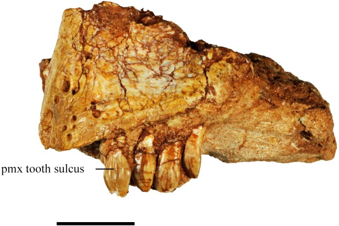
Material from a minimum of twenty-nine individuals of a new ornithopod, represented by nearly every skeletal element, was recovered from the Proctor Lake locality in the Twin Mountains Formation (Aptian) of north-central Texas. This material includes various ontogenetic stages, providing insight into the growth patterns of this species. The new ornithopod, Convolosaurus marri gen. et sp. nov., is recovered outside of Iguanodontia, but forms a clade with Iguanodontia exclusive of Hypsilophodon foxii. The presence and morphology of four premaxillary teeth along with a combination of both basal and derived characters distinguish this taxon from all other ornithopods. Basal characters present in C. marri including the presence of premaxillary teeth, the shape of the dentary teeth, and position of the pterygoid wing on the quadrate, whereas the presence of opisthocoelous cervical vertebrae, large proximal caudal neural spines, and curved maxillary tooth roots suggest C. marri is more derived than 80% of the basal neornithischians included in this analysis.


Conflict of interest statement

The authors have declared that no competing interests exist.

Figures

Fig 1
Fig 1. Map of study area.
Map of Proctor Lake, Texas located in Comanche County (green) with marked locations of the two quarries (Camp Quarry and North Quarry) where ornithopod fossils were collected.
Fig 2
Fig 2. Articulated specimens.
(A) SMU 70456, articulated subadult individual on display at the Proctor Lake Corps of Engineers Office. Scale arrow equals 10 cm. (B) Composite skeleton on display at the Perot Museum of Nature and Science. Scale bar equals 10 cm. (C) SMU 75379 and SMU 75380, partial articulated skeletons found stacked on one another. Scale bar equals 5 cm.
Fig 3
Fig 3. Modified stratigraphic column of Proctor Lake fossil locality.
Stratigraphic section of Twin Mountains Formation exposed at Proctor Lake (modified from Winkler and Murry, 1989).
Fig 4
Fig 4. Skull reconstruction.
Skull reconstruction of C. marri based on available specimens. Abbreviations: A-articular, BO-basioccipital, D-dentary, F-frontal, J-jugal, L-lacrimal, MX-maxilla, N-nasal, OP-opisthotic, P-parietal, PD-predentary, PF-prefrontal, PMX-premaxilla, PO-postorbital, Q-quadrate, QJ-quadratojugal, SA-surangular, SOB-supraorbital, SQ-squamosal.
Fig 5
Fig 5. Premaxilla from specimen SMU 72316.
Left premaxilla (SMU 72316) in lateral view containing a rugose anterodorsal surface and four premaxillary teeth displaying basal-apical oriented sulcus on the premaxillary teeth. Scale bar equals 1 cm.
Fig 6
Fig 6. SMU 72834 anterior skull.
(A) SMU 72834, anterior skull in right lateral view. (B) Illustration of SMU 72834, in right lateral view (David Baker). Abbreviations: D-dentary, L-lacrimal, MX-maxilla, PMX-premaxilla, PD-predentary, QJ-quadratojugal. Scale bar equals 5 cm.
Fig 7
Fig 7. SMU 74749 and SMU 72316 cranial material.
(A) SMU 74749, partial cranium in right lateral view. (B) Outline drawing of identifiable bone contacts on the right side of SMU 74749 partial cranium. (C) SMU 72316, partial cranium in right lateral view. (D). Outline drawing of identifiable bone contacts on the right side of SMU 72316 partial cranium. Abbreviations: D-dentary, F-frontal, J-jugal, MX-maxilla, MX proc.-anterolateral process of the maxilla, N-nasal, PF-prefrontal, PMX-premaxilla, PMX fo- premaxillary foramen. Scale bar equals 5 cm.
Fig 8
Fig 8. SMU 74678 cranium.
(A) SMU 74678, posterior skull in right lateral view. This specimen contains two supraorbitals which are not fused to the frontal. (B) Outline drawing of right side of SMU 74678 posterior skull. Abbreviations: F-frontal, J-jugal, L-lacrimal, N-nasal, P-parietal, PF-prefrontal, PO-postorbital, Q-quadrate, QJ- quadratojugal, QJ fo- quadratojugal foramen, SOB-supraorbital, SQ-squamosal. Scale bar equals 5 cm.
Fig 9
Fig 9. SMU 72834 partial cranium.
(A) SMU 72834, posterior cranium in right lateral view. (B) SMU 72834, posterior cranium in posterior view. (C) SMU 72834, posterior cranium in dorsal view. (D) Outline drawing of dorsal view of SMU 72834. Abbreviations: BSP proc- basipterygoid process, F-frontal, P-parietal, PO-postorbital, SOB-supraorbital, SQ-squamosal, Q-quadrate. Scale bar equals 5 cm.
Fig 10
Fig 10. Occipital region of SMU 72834.
(A) SMU 72834, occipital region in ventral view. (B) Outline drawing of SMU 72834, occipital region in ventral view. (C) SMU 71504, right distal paroccipital process in ventral view. (D) SMU 71504, right distal paroccipital process in dorsal view. Abbreviations: BO-basioccipital, EO-exoccipital, OP-opisthotic, P-parietal, SO-supraoccipital. Scale bar equals 5 cm.
Fig 11
Fig 11. SMU 72316 Maxillary and dentary teeth.
(A) SMU 72316, maxillary tooth lateral view. (B) SMU 72316, maxillary tooth medial view. (C) SMU 72316, dentary tooth medial view. (D). SMU 72316, dentary tooth lateral view. Scale bar equals 1 cm.
Fig 12
Fig 12. SMU 75621 axis and third cervical vertebra.
Axis and third cervical vertebrae in left lateral view. Abbreviations: d-diapophysis, p-parapophysis. Scale bar equals 1 cm.
Fig 13
Fig 13. SMU 72834 cervical vertebrae.
(A) Dorsal view of cervical vertebrae 4–9. (B) Left lateral view of cervical vertebrae 4–9. (C) Ventral view of cervical vertebrae 4–9. Abbreviations: d-diapophysis, ns-neural spine, p-parapophysis, prz-prezygapophyses, poz-postzygapophyses. Scale bar equals 5 cm.
Fig 14
Fig 14. SMU 72316 dorsal vertebrae.
(A) Left lateral view of dorsal vertebrae nine through 15. (B) Ventral view of dorsal vertebrae nine through 15. Scale bar equals 3 cm.
Fig 15
Fig 15. SMU 72316 pelvic girdle.
(A) SMU 72316, pelvic girdle left lateral view. (B) SMU 72316, pelvic girdle ventral view. Scale bar equals 5 cm.
Fig 16
Fig 16. Caudal vertebrae.
(A) SMU 70456, proximal caudal vertebra in left lateral view. (B) SMU 71492, caudal centra in dorsal view displaying anteroposteriorly elongate foramen. (C) SMU 72316, distal caudal vertebrae (#32–34) in left lateral view. Abbreviations: fo-foramen. Scale bar equals 3 cm.
Fig 17
Fig 17. Ossified tendons in the caudal region.
(A) SMU 74670, caudal vertebrae with ossified tendons preserved in right lateral view. (B) Outline drawing of SMU 74670, caudal vertebrae and ossified tendons. (C) SMU 72316, articulated caudal vertebrae (#16–26) with ossified tendons preserved in left lateral view. Abbreviations: ns-neural spine. Scale bar equals 5 cm.
Fig 18
Fig 18. SMU 72834 scapula and coracoid.
SMU 75564 humerus. (A) SMU 72834, right scapula in lateral view. (B) SMU 72834, right scapula in medial view. (C) SMU 72834, right scapula in dorsal view. (D) SMU 72834, right coracoid in medial view. (E) SMU 72834, right coracoid in lateral view. (F) SMU 75564, right humerus in posterior view. (G) SMU 75564, right humerus in lateral view. (H) SMU 75564, right humerus in medial view. Abbreviations: co fo-coracoid foramen, co sul- coracoid sulcus, gl-glenoid, sc dep- scapular depression, sc ridge- scapular ridge, sc spine-scapular spine, delt-p.c.-delta pectoral crest, fos-fossa, r. cond.-radius condyle, u cond.-ulna condyle. Scale bar equals 5 cm.
Fig 19
Fig 19. SMU 71821 ulna and SMU 71818 radius.
(A) SMU 71821, left ulna in lateral view. (B) SMU 71821, left ulna in medial view. (C) SMU 71821, left ulna in posterior view. (D) SMU 71821, left ulna in anterior view. (E) SMU 71821, left ulna in proximal view. (F) SMU 71821, left ulna in distal view. (G) SMU 71818, left radius in lateral view. (H) SMU 71818, left radius in medial view. (I) SMU 71818, left radius in posterior view. (J) SMU 71818, left radius in anterior view. (K) SMU 71818, left radius in proximal view. (L) SMU 71818, left radius in distal view. Scale bar equals 2 cm.
Fig 20
Fig 20. SMU 70456 left manus.
Left manus (SMU 70456) in dorsal view. Abbreviations: I, II, III, IV, V- digits one through five, Mc-metacarpals, R-radius, U-ulna. Scale bar equals 5 cm.
Fig 21
Fig 21. SMU 77617 left ilium.
(A) Left ilium (SMU 77617) in lateral view. (B) Left ilium in medial view. (C) Left ilium in dorsal view with anterior end to the left. Abbreviations: acet.-acetabulum, isp- ischial peduncle, pp- pubic peduncle, prac- preacetabular process. Scale bar equals 5 cm.
Fig 22
Fig 22. Ischium and pubis.
(A) SMU 74119, right ischium in lateral view. (B) SMU 74119, right ischium in medial view. (C). SMU 74119, right ischium in ventral view. (D) SMU 77638, distal left ischium lateral view. (E) SMU 77638, distal left ischium dorsal view. (F) SMU 74679, left partial pubis in lateral view. (G) SMU 72316, left partial pubis. (H) SMU 75636, left partial pubis. Abbreviations: ip- iliac peduncle, obt. proc.- obturator process, open fo-open foramen, pp-pubic peduncle, prepubic proc.-prepubic process. Scale bar 5 cm.
Fig 23
Fig 23. Femora.
(A) SMU 74665, right femur in posterior view. (B) SMU 74665, right femur in medial view. (C). SMU 74665, right femur in anterior view. (D) SMU 72451, left femur ventral view. (E) SMU 70456, left femur ventral view. Abbreviations: gr. troch.- greater trochanter, lc-lateral condyle, les. troch.- lesser trochanter, mc-medial condyle, 4th troch.- fourth trochanter. Scale bar 5 cm.
Fig 24
Fig 24. Hind limb.
(A) SMU 72316, left hind limb in lateral view. (B) SMU 72316, left hind limb in medial view. (C). SMU 72316, left hind limb in posterior view. (D) SMU 77617, right tibia, fibula, astragalus, calcaneum in lateral view. (E) SMU 77617, right tibia, fibula, astragalus, calcaneum in posterior view. (F) SMU 77617, right tibia, fibula, astragalus, calcaneum in medial view. (G) SMU 77617, right tibia in anterior view. (H) SMU 77617, right tibia in ventral view. (I) SMU 72834, proximal left tibia in dorsal view. Abbreviations: As- astragalus, Ca- calcaneum, cnem. c- cnemial crest, Fem-femur, Fib- fibula, lc-lateral condyle, mc-medial condyle, Tib-tibia, 4th troch.- fourth trochanter. Scale bar 5 cm.
Fig 25
Fig 25. Astragalus and calcaneum.
(A) SMU 72534, left calcaneum in medial view. (B) SMU 72534, left calcaneum in lateral view. (C). SMU 72534, left calcaneum in anterior view. (D) SMU 71690, left astragalus in medial view. (E) SMU 71690, left astragalus in lateral view. (F) SMU 71690, left astragalus in anterior view. (G) SMU 71690, left astragalus in posterior view. (H) SMU 71690, left astragalus in dorsal view. (I) SMU 71690, left astragalus in ventral view. Abbreviations: Asc proc- astragalus ascending process, fos-fossa. Scale bar 1 cm.
Fig 26
Fig 26. Pes.
(A) SMU 77636, left articulated pes in dorsal view. (B) SMU 77636, left articulated metatarsals in proximal view. Abbreviations: I, II, III, IV- digits one through four. Scale bar equals 3 cm.
Fig 27
Fig 27. Femur size distribution from Proctor Lake.
Scatter plot of femur length versus proximal and distal width from Proctor Lake. Nineteen complete femora were used to create two linear regression models which was then used to estimate the length of 26 additional partial femora based on proximal and distal width. Measurements listed in S1 Table.
Fig 28
Fig 28. Histogram of femur length distribution from Proctor Lake fossil locality.
Histogram of femur length distribution from Proctor Lake fossil locality. Graph displaying range of femoral length of 29 individuals recovered from the Proctor Lake locality.
Fig 29
Fig 29. Histogram of femur length distribution from Proctor Lake locality fossil sites.
Graph displaying range of femoral length of individuals found in clusters. Site localities are listed along the horizontal axis.
Fig 30
Fig 30. Strict consensus tree produced from phylogenetic analysis.
Strict consensus tree of 96 most parsimonious trees recovered from phylogenetic analysis. Bootstrap support values >50% listed beneath nodes.

References

    1. Winkler DA, Murry PA. Paleoecology and hypsilophodontid behavior at the Proctor Lake dinosaur locality (Early Cretaceous), Texas. Geological Society of America Special Paper. 1989;238: 55–61.
    1. Young K. Comanche Series (Cretaceous), south central Texas. Permian Basin Section, Society of Economic Paleontologists and Mineralogists. 1967;9–29.
    1. Winkler DA, Murry PA, Jacobs LL. Early Cretaceous (Comanchean) Vertebrates of Central Texas. Journal of Vertebrate Paleontology. 1990;10: 95–116.
    1. Jacobs LL, Winkler DA. Mammals, archosaurs, and the Early to Late Cretaceous transition in north-central Texas 1998;253–280 in Tomida Y, Flynn L.J., Jacobs L.L., editors. Advances in vertebrate paleontology and geochronology. National Science Museum, Tokyo.
    1. Tanrikulu S, Doyle JA, Delusina I. Early Cretaceous (Albian) spores and pollen from the Glen Rose Formation of Texas and their significance for correlation of the Potomac Group. Palynology. 2017;1–19.

Publication types