

Quantitative and semi-quantitative computed tomography analysis of interstitial lung disease associated with systemic sclerosis: A longitudinal evaluation of pulmonary parenchyma and vessels
- PMID: 30861018
- PMCID: PMC6414027
- DOI: 10.1371/journal.pone.0213444
Quantitative and semi-quantitative computed tomography analysis of interstitial lung disease associated with systemic sclerosis: A longitudinal evaluation of pulmonary parenchyma and vessels
Abstract
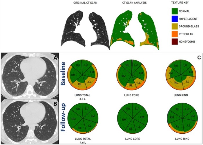
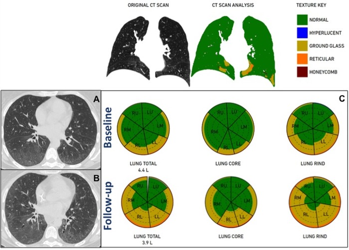
Objectives: To evaluate interstitial lung disease associated with systemic sclerosis (SSc-ILD) and its changes during treatment by using quantitative analysis (QA) compared to semi-quantitative analysis (semiQA) of chest computed tomography (CT) scans. To assess the prognostic value of QA in predicting functional changes.
Materials and methods: We retrospectively selected 35 consecutive patients with SSc-ILD with complete pulmonary functional evaluation, Doppler-echocardiography, immunological tests, and chest CT scan at both baseline and follow-up after immunosuppressive therapy. CT images were analyzed by two chest radiologists for semiQA and by a computational platform for texture analysis of ILD patterns (CALIPER) for QA. Concordance between semiQA and QA was tested. Traction bronchiectasis severity was scored. Analysis of ROC curves was performed.
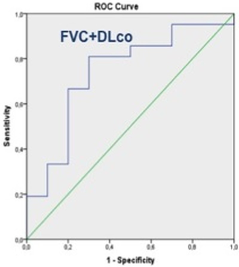
Results: Seventy CT scans were analyzed and QA failed in 4/70 scans. Thus, the final population included 31/35 patients (51.3±12.1 years). QA had a weak-to-good concordance with semiQA (ICC reticular:0.275; ICC ground-glass:0.667) and QA correlated better than semiQA (r = -0.3 to -0.74 vs r = -0.3 to -0.4) with functional parameters. Both methods correlated with traction bronchiectases score and pulmonary artery diameter at CT. A pulmonary artery diameter ≥29mm distinguished patients with lower lung volumes and ILD extent greater than 39% (p<0.001). Changes in QA patterns during treatment were not accurate (AUC: 0.50 to 0.70; p>0.05) in predicting disease progression as assessed by functional parameters, whereas variation in total lung volume at QA accurately predicted changes in the composite functional respiratory endpoint with FVC% and DLco% (AUC = 0.74; 95%CI: 0.54 to 0.93; p = 0.03).
Conclusions: Pulmonary QA of CT images can objectively quantify specific patterns of ILD changes during treatment in patients with SSc-ILD. Changes in QA patterns do not correlate with functional changes, but variation in total lung volume at QA accurately predicted changes in the composite functional respiratory endpoint with FVC% and DLco%. Pulmonary artery diameter at CT reflects the interstitial involvement, identifying patients with more severe prognosis.
Conflict of interest statement
MO received consultancies from Imbio, LLC, all remaining authors have declared that no competing interests exist. This does not alter our adherence to PLOS ONE policies on sharing data and materials.
Figures
References
-
- Steen VD, Owens GR, Fino GJ, Rodnan GP, Medsger TA Jr. Pulmonary involvement in systemic sclerosis (scleroderma). Arthritis Rheum 1985; 28:759–767. - PubMed
-
- Desai SR, Veeraraghavan S, Hansell DM, Nikolakopolou A, Goh NS, Nicholson AG, et al. CT features of lung disease in patients with systemic sclerosis: comparison with idiopathic pulmonary fibrosis and nonspecific interstitial pneumonia. Radiology 2004; 232:560–567. - PubMed
MeSH terms
Substances
LinkOut - more resources
Full Text Sources
Medical
