. 2019 Oct 1;527(14):2291-2301.
doi: 10.1002/cne.24675. Epub 2019 Mar 25.

Absence of axonal sprouting following unilateral lesion in 125-day-old rat supraoptic nucleus may be due to age-dependent decrease in protein levels of ciliary neurotrophic factor receptor alpha

Affiliations

Absence of axonal sprouting following unilateral lesion in 125-day-old rat supraoptic nucleus may be due to age-dependent decrease in protein levels of ciliary neurotrophic factor receptor alpha

Jason M Askvig et al. J Comp Neurol. .

Abstract

Within the supraoptic nucleus (SON) of a 35-day-old rat, we previously demonstrated a collateral sprouting response that reinnervates the partially denervated neural lobe (NL) after unilateral lesion of the hypothalamo-neurohypophysial tract. Others have shown a decreased propensity for axonal sprouting in an aged brain; therefore, to see if the SON exhibits a decreased propensity for axonal sprouting as the animal ages, we performed a unilateral lesion in the 125-day-old rat SON. Ultrastructural analysis of axon profiles in the NL of the 125-day-old rat demonstrated an absence of axonal sprouting following injury. We previously demonstrated that ciliary neurotrophic factor (CNTF) promotes process outgrowth from injured magnocellular neuron axons in vitro. Thus, we hypothesized that the lack of axonal sprouting in the 125-day-old rat SON may be due to a reduction in CNTF or the CNTF receptor components. To this point, we found that as the rat ages there is significantly less CNTF receptor alpha (CNTFRα) protein in the uninjured, 125-day-old rat compared to the uninjured, 35-day-old rat. We also observed that protein levels of CNTF and the CNTF receptor components were increased in the SON and NL following injury in the 35-day-old rat, but there was no difference in the protein levels in the 125-day-old rat. Altogether, the results presented herein demonstrate that the plasticity within the SON is highly dependent on the age of the rat, and that a decrease in CNTFRα protein levels in the 125-day-old rat may contribute to the loss of axonal sprouting following axotomy.

Keywords: CNTF; CNTFRα; RRID:AB_2136105; RRID:AB_2276485; RRID:AB_397120; RRID:AB_476697; RRID:AB_518526; RRID:AB_518680; RRID:AB_631590; aging; axonal sprouting; neural lobe; supraoptic nucleus.


Figures

Figure 1.
Figure 1.
Failure of lesion-induced axonal sprouting in 125-day-old rat. Following unilateral lesion of the hypothalamo-neurohypophysial tract, the number of axons is significantly decreased at one-week post-surgery compared to intact and sham-control (p<0.0001). Moreover, the number of axons remains significantly decreased in the lesion NL at four-weeks post-surgery compared to intact and sham-control (p<0.0001).
Figure 2.
Figure 2.
Urine osmolarity remains reduced following unilateral lesion in the 125-day-old rat. Following unilateral lesion, urine osmolality in the 125-day-old rat was significantly decreased (p<0.01) compared to the response following unilateral lesion in the 35-day-old rat. The decrease is first observed two-days post-surgery and persisted throughout the entire three-week post-surgery period. The elevated urine osmolarity in the 35-day-old rat post-lesion is indicative of heightened neurosecretory activity of the magnocellular neurons.
Figure 3.
Figure 3.
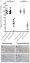
Unilateral lesion results in less oxytocinergic and vasopressinergic immunoreactive profiles in the 125-day SON. Cell counts demonstrated no significant difference in the number of immunoreactive profiles indicative of oxytocinergic (p=0.6941) or vasopressinergic (p=0.9851) neurons in the SON contralateral to the unilateral hypothalamic lesion. However, at 10 dpl the numbers of immunoreactive profiles indicative of oxytocinergic and vasopressinergic neurons were reduced by 88% and 93%, respectively, in the lesion SON (a). Representative images demonstrate the decrease in oxytocinergic neurons between uninjured control (b) and lesioned SON (c) and the decrease in vasopressinergic neurons between uninjured control (d) and lesioned SON (e). The oxytocin- and vasopressin-immunoreactive profiles in b-e are in brown while the purple represents the cresyl violet-labeled nucleoli. Each shape on the graph indicates an individual data point with the lines representing the mean and SD. Each data point is comprised of a minimum of six sections sampled from each of [n] animals. The SON is outlined by the dashed line. OC = optic chiasm. Magnification bar = 100μm. ***p<0.0001.
Figure 4.
Figure 4.
Illustration of CNTF and CNTF receptor complex. CNTF requires three signaling molecules to transduce the signaling response intracellularly, CNTFRα, LIFRß, and gp130. CNTFRα is GPI-anchored to the extracellular membrane while LIFRß and gp130 are transmembrane receptors. The LIFRß and gp130 receptor molecules are not associated with CNTFRα until CNTF binds to CNTFRα (a). When CNTF binds CNTFRα, it causes a translocation of LIFRß and gp130 to interact with CNTFRα and LIFRß and gp130 are then able to transduce the signal intracellularly (b). Images created with BioRender.
Figure 5.
Figure 5.
CNTFRα protein levels decreased in the uninjured SON with age. Western blot analysis revealed a significant decrease in CNTFRα protein levels in the control SON from 35- to 125-days of age (a), but no change in CNTFRα protein levels in the maturing NL (c; p=0.2978). Analysis revealed no change in protein levels of CNTF in the maturing SON (b; p=0.07) or maturing NL (c; p=0.16), LIFRß in the maturing SON (b; p=0.10) or maturing NL (c; p=0.27), or gp130 in the maturing SON (b; p=0.28) or maturing NL (c; p=0.15). Each shape on the graph indicates an individual data point with the lines representing the mean and SD. Each data point represents isolated SON pooled from six rats with all experiments repeated in triplicate. *p<0.05, **p<0.01
Figure 6.
Figure 6.
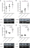
Unilateral lesion results in increased levels of CNTF and CNTF receptor complex proteins in the 125-day-rat SON. Western blot analysis demonstrated a significant increase in CNTF (a; p=0.014), CNTFRα (b; p=0.0013), LIFRß (c; p=0.04), and gp130 (d; p<0.0001) protein levels in the injured (lesion) SON at 10 dpl. However, there was no significant difference in the protein levels in the SON contralateral to injury compared to age-matched, uninjured, control for CNTF (a; p=0.23), CNTFRα (b; p=0.075), LIFRß (c; p=0.07), and gp130 (d; p=0.36). Each shape on the graph indicates an individual data point with the lines representing the mean and SD. Each data point represents isolated SON pooled from six rats with all experiments repeated in triplicate. *p<0.05, **p<0.01, ***p<0.0001.
Figure 7.
Figure 7.
Protein levels of CNTF and CNTF receptor complex increase in the 35-day rat NL following unilateral lesion. Western blot analysis demonstrated a significant increase in CNTF (a; p=0.002), CNTFRα (b; p<0.0001), LIFRß (c; p=0.0003), and gp130 (d; p=0.018) protein levels in the lesion NL compared to age-matched control. Each shape on the graph indicates an individual data point with the lines representing the mean and SD. Each data point represents isolated SON pooled from six rats with all experiments repeated in triplicate. *p<0.05, **p<0.01, ***p<0.0001.
Figure 8.
Figure 8.
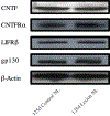
Protein levels of CNTF and CNTF receptor complex did not change in the 125-day rat NL following unilateral lesion. Western blot analysis demonstrated no significant difference in CNTF (p=0.68), CNTFRα (p=0.24), LIFRß (p=0.45), and gp130 (p=0.99) protein levels in the 125 day lesion NL compared to age-matched control. Images are representative protein bands with all experiments repeated in triplicate.

References

    1. Adams JH, Daniel PM, & Prichard MM (1964). Some Effects of Transection of the Pituitary Stalk. British Medical Journal, 2(5425), 1619–1625. - PMC - PubMed
    1. Armstrong WE, & Hatton GI (1978). Morphological changes in the rat supraoptic and paraventricular nuclei during the diurnal cycle. Brain Research, 157(2), 407. - PubMed
    1. Askvig JM, Leiphon LJ, & Watt JA (2012). Neuronal activity and axonal sprouting differentially regulate CNTF and CNTF receptor complex in the rat supraoptic nucleus. Experimental Neurology, 233(1), 243. - PMC - PubMed
    1. Askvig JM, Lo DY, Sudbeck AW, Behm KE, Leiphon LJ, & Watt JA (2013). Inhibition of the Jak-STAT pathway prevents CNTF-mediated survival of axotomized oxytocinergic magnocellular neurons in organotypic cultures of the rat supraoptic nucleus. Experimental Neurology, 240, 75. - PMC - PubMed
    1. Askvig JM, & Watt JA (2015). The MAPK and PI3K pathways mediate CNTF-induced neuronal survival and process outgrowth in hypothalamic organotypic cultures. Journal of Cell Communication and Signaling, 9(3), 217. - PMC - PubMed

Publication types

Substances