Myositis Autoantigen Expression Correlates With Muscle Regeneration but Not Autoantibody Specificity
- PMID: 30861336
- PMCID: PMC6663619
- DOI: 10.1002/art.40883
Myositis Autoantigen Expression Correlates With Muscle Regeneration but Not Autoantibody Specificity
Abstract
Objective: Although more than a dozen myositis-specific autoantibodies (MSAs) have been identified, most patients with myositis are positive for a single MSA. The specific overexpression of a given myositis autoantigen in myositis muscle has been proposed as initiating and/or propagating autoimmunity against that particular autoantigen. The present study was undertaken to test this hypothesis.
Methods: In order to quantify autoantigen RNA expression, RNA sequencing was performed on muscle biopsy samples from control subjects, MSA-positive patients with myositis, regenerating mouse muscles, and cultured human muscle cells.
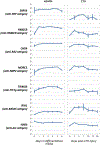
Results: Muscle biopsy samples were available from 20 control subjects and 106 patients with autoantibodies recognizing hydroxymethylglutaryl-coenzyme A reductase (n = 40), signal recognition particles (n = 9), Jo-1 (n = 18), nuclear matrix protein 2 (n = 12), Mi-2 (n = 11), transcription intermediary factor 1γ (n = 11), or melanoma differentiation-associated protein 5 (n = 5). The increased expression of a given autoantigen in myositis muscle was not associated with autoantibodies recognizing that autoantigen (all q > 0.05). In biopsy specimens from both myositis muscle and regenerating mouse muscles, autoantigen expression correlated directly with the expression of muscle regeneration markers and correlated inversely with the expression of genes encoding mature muscle proteins. Myositis autoantigens were also expressed at high levels in cultured human muscle cells.
Conclusion: Most myositis autoantigens are highly expressed during muscle regeneration, which may relate to the propagation of autoimmunity. However, factors other than overexpression of specific autoantigens are likely to govern the development of unique autoantibodies in individual patients with myositis.
© 2019 American College of Rheumatology. This article has been contributed to by US Government employees and their work is in the public domain in the USA.
Conflict of interest statement
Figures


Similar articles
-
Autoantibodies in polymyositis and dermatomyositis.Curr Rheumatol Rep. 2013 Jun;15(6):335. doi: 10.1007/s11926-013-0335-1. Curr Rheumatol Rep. 2013. PMID: 23591825 Review.
-
Autoantibodies against 3-hydroxy-3-methylglutaryl-coenzyme A reductase in patients with statin-associated autoimmune myopathy.Arthritis Rheum. 2011 Mar;63(3):713-21. doi: 10.1002/art.30156. Arthritis Rheum. 2011. PMID: 21360500 Free PMC article.
-
Cutting edge issues in polymyositis.Clin Rev Allergy Immunol. 2011 Oct;41(2):179-89. doi: 10.1007/s12016-010-8238-7. Clin Rev Allergy Immunol. 2011. PMID: 21191666 Review.
-
Pathological autoantibody internalisation in myositis.Ann Rheum Dis. 2024 Oct 21;83(11):1549-1560. doi: 10.1136/ard-2024-225773. Ann Rheum Dis. 2024. PMID: 38902010
-
Enhanced autoantigen expression in regenerating muscle cells in idiopathic inflammatory myopathy.J Exp Med. 2005 Feb 21;201(4):591-601. doi: 10.1084/jem.20041367. J Exp Med. 2005. PMID: 15728237 Free PMC article.
Cited by
-
Association of anti-HMGCR antibodies of the IgM isotype with refractory immune-mediated necrotizing myopathy.Arthritis Res Ther. 2024 Sep 11;26(1):158. doi: 10.1186/s13075-024-03387-6. Arthritis Res Ther. 2024. PMID: 39261921 Free PMC article.
-
Immune-mediated necrotizing myopathy: clinical features and pathogenesis.Nat Rev Rheumatol. 2020 Dec;16(12):689-701. doi: 10.1038/s41584-020-00515-9. Epub 2020 Oct 22. Nat Rev Rheumatol. 2020. PMID: 33093664 Review.
-
Coordinated local RNA overexpression of complement induced by interferon gamma in myositis.Sci Rep. 2023 Feb 4;13(1):2038. doi: 10.1038/s41598-023-28838-z. Sci Rep. 2023. PMID: 36739295 Free PMC article.
-
Distinct Cytokine and Cytokine Receptor Expression Patterns Characterize Different Forms of Myositis.medRxiv [Preprint]. 2025 Feb 21:2025.02.17.25321047. doi: 10.1101/2025.02.17.25321047. medRxiv. 2025. Update in: Rheumatology (Oxford). 2025 Jun 20:keaf346. doi: 10.1093/rheumatology/keaf346. PMID: 40034760 Free PMC article. Updated. Preprint.
-
Variants in DTNA cause a mild, dominantly inherited muscular dystrophy.Acta Neuropathol. 2023 Apr;145(4):479-496. doi: 10.1007/s00401-023-02551-7. Epub 2023 Feb 17. Acta Neuropathol. 2023. PMID: 36799992 Free PMC article.
References
-
- Dalakas MC. Inflammatory Muscle Diseases. N Engl J Med 2015;373:393–4. - PubMed
Publication types
MeSH terms
Substances
Grants and funding
LinkOut - more resources
Full Text Sources
Medical

