. 2019 May;119(5):744-757.
doi: 10.1055/s-0039-1679908. Epub 2019 Mar 12.

Epithelial (E)-Cadherin is a Novel Mediator of Platelet Aggregation and Clot Stability

Affiliations

Epithelial (E)-Cadherin is a Novel Mediator of Platelet Aggregation and Clot Stability

Vanessa M Scanlon et al. Thromb Haemost. 2019 May.

Abstract

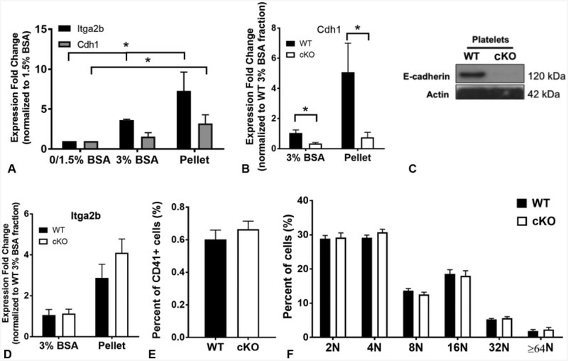
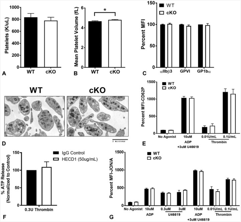
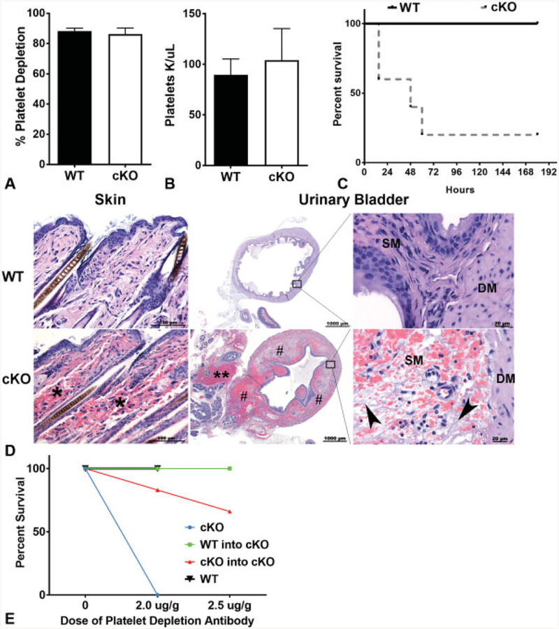
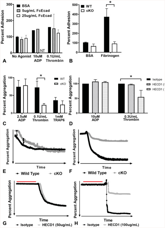
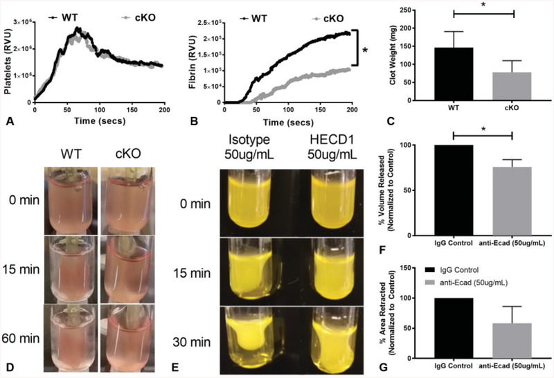
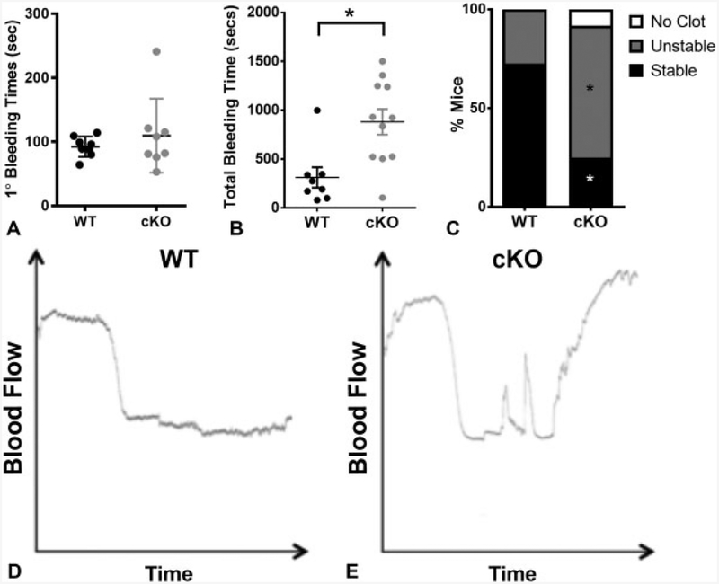
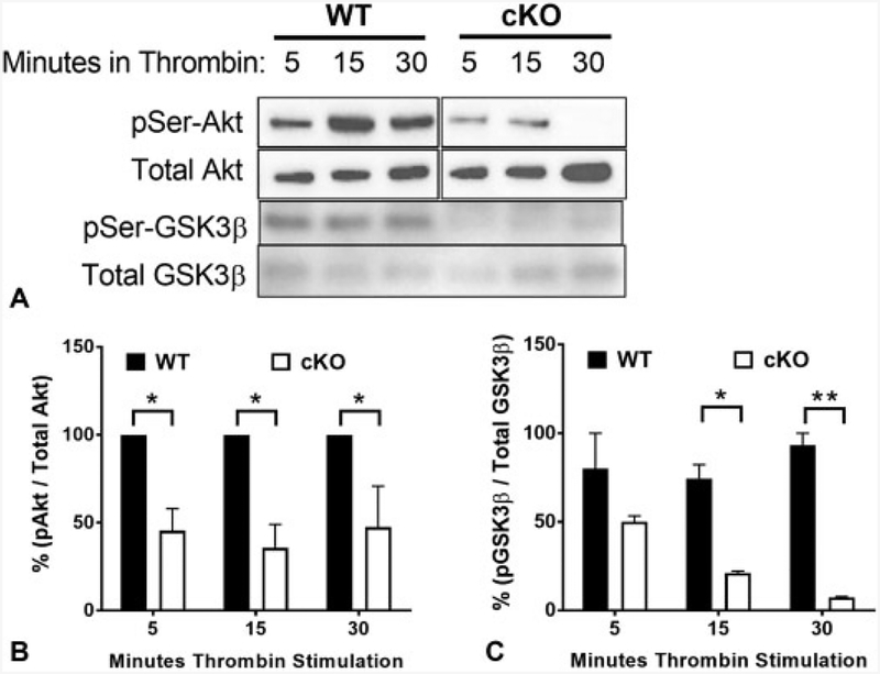
Cadherins play a major role in mediating cell-cell adhesion, which shares many parallels with platelet-platelet interactions during aggregate formation and clot stabilization. Platelets express epithelial (E)-cadherin, but its contribution to platelet function and/or platelet production is currently unknown. To assess the role of E-cadherin in platelet production and function in vitro and in vivo, we utilized a megakaryocyte-specific E-cadherin knockout mouse model. Loss of E-cadherin in megakaryocytes does not affect megakaryocyte maturation, platelet number or size. However, platelet dysfunction in the absence of E-cadherin is revealed when conditional knockout mice are challenged with acute antibody-mediated platelet depletion. Unlike wild-type mice that recover fully, knockout mice die within 72 hours post-antibody administration, likely from haemorrhage. Furthermore, conditional knockout mice have prolonged tail bleeding times, unstable clot formation, reduced clot retraction and reduced fibrin deposition in in vivo injury models. Murine platelet aggregation in vitro in response to thrombin and thrombin receptor activating peptide is compromised in E-cadherin null platelets, while aggregation in response to adenosine diphosphate (ADP) is not significantly different. Consistent with this, in vitro aggregation of primary human platelets in response to thrombin is decreased by an inhibitory E-cadherin antibody. Integrin activation and granule secretion in response to ADP and thrombin are not affected in E-cadherin null platelets, but Akt and glycogen synthase kinase 3β (GSK3β) activation are attenuated, suggesting a that E-cadherin contributes to aggregation, clot stabilization and retraction that is mediated by phosphoinositide 3-kinase/Akt/GSK3β signalling. In summary, E-cadherin plays a salient role in platelet aggregation and clot stability.


Conflict of interest statement

None declared.

Figures

Fig. 1
Fig. 1
Epithelial (E)-cadherin is expressed in murine megakaryocytes. (A) Relative messenger ribonucleic acid (mRNA) expression in bovine serum albumin (BSA) gradient sub-fractions following in vitro differentiation of wild-type (WT) foetal liver cells. Itga2b (positive control) expression is significantly higher in the 3% and pellet fractions than the 0/1.5% BSA fraction (p < 0.05), and Cdh1 is significantly higher in the pellet fraction (p < 0.05). (B)Cdh1 mRNAis significantly reduced in differentiated foetal liver cells isolated from E-cadherin conditional knockout (cKO) embryos (p < 0.05). Data are presented as fold change normalized to the WT 3% BSA fraction ± standard deviation (SD) (n = 3). (C) Cropped Western blot of isolated murine platelets from WT and cKO mice demonstrating loss of E-cadherin protein in cKO platelet lysates. The blot was cut at 75 kDa, and the top portion blotted for E-cadherin, the bottom portion blotted for actin. This blot is representative of 5 repeats. (D) mRNA expression of Itga2b in BSA gradient sub-fractions following in vitro differentiation of foetal liver cells isolated from WT and cKO embryos. Data are presented as fold change normalized to the 3% BSA fraction ± SD (n = 3). (E) Flow cytometry on murine bone marrow (BM) of total per cent CD41+ cells, and (F) ploidy of BM-derived megakaryocytes (WT n = 5, cKO n = 4).
Fig. 2
Fig. 2
Thrombocytopoiesis and granule secretion are not affected by epithelial (E)-cadherin. (A) Platelet counts in wild-type (WT) (n = 12) and conditional knockout (cKO) (n = 20) animals. Average absolute values ± standard deviation (SD) are shown. (B) Mean platelet volume between WT (n = 12) and cKO (n = 20) animals. There is a small, but significant increase in mean platelet volume in cKO mice (p < 0.05). Average absolute values ± SD are represented. (C) Cell surface expression by flow cytometry of platelet-critical in tegrins and glycoproteins. Data are presented as per cent mean fluorescence intensity (MFI) normalized to WT ± SD(n ≥ 6). (D) Electron microscopy images of resting platelets from WT and cKO mice show no obvious differences in granule number or distribution. (E) Alpha granule release in response to indicated agonists was measured by cell surface expression of P-selectin (CD62P). Data are presented as percent MFI normalized to WT without agonist ± SD (n ≥ 7). (F) Dense granule release in human platelets inhibited with an antibody against E-cadherin (HECD1) in response to thrombin was measured by cell adenosine triphosphate (ATP) release. Data are presented as per cent ATP release normalized to control-treated platelets ± SD(n = 3). (G) Washed platelets were stim ulated with the indicated concentrations of adenosine diphosphate (ADP), U46619 or thrombin and activation assessed via activated αIIbβ3 (JON/A). Data are presented as per cent MFI normalized to WT without agonist ± SD (n ≥ 6).
Fig. 3
Fig. 3
Epithelial (E)-cadherin conditional knockout (cKO) animals are sensitive to bleeding and death in response to immune-mediated thrombocytopenia. (A) Percent platelet depletion in wild-type (WT) and cKO mice after 6 to 10 hours of treatment with anti-CD42b (2 mg/g) (n ≥ 5). Data are presented as mean ± standard deviation (SD). (B) Absolute number of platelets remaining 6 to 10 hours after depletion with anti-CD42b (2 mg/g) between WT and cKO mice (n ≥ 5). Data are presented as mean ± SD. (C) Survival of animals following platelet depletion (n ≥ 5, p < 0.01). (D) Representative photomicrographs of haematoxylin and eosin (H&E)-stained sections of skin and urinary bladder (low and high magnification) in WT and cKO mice 7 hours post-antibody treatment. There is multi-focal marked dermal haemorrhage (*), massive haemorrhage within the adventitia (**) adjacent to the urinary bladder and marked and multi-focal haemorrhage (#) within the urinary bladder submucosa and detrusor muscle in cKO mice compared with WT mice. Higher power images of the boarder of the submucosa (SM) and detrusor muscle (DM) reveals there is fibrin (pale fibrillary material, arrow heads), haemorrhage and oedema (clear spaces) within submucosa of cKO mice compared with WT mice. (E) Survival of animals following platelet depletion with increasing doses of anti-CD42b in transplanted and untransplanted animals. cKO: untransplanted cKO mice (n = 3); WT: untransplanted WT mice (n = 3); WT-into-cKO: cKO mice transplanted with WT bone marrow (n = 5); cKO-into-cKO: cKO mice transplanted with cKO bone marrow (n = 6).
Fig. 4
Fig. 4
Fibrinogen binding is affected, and aggregation is impaired in the absence of epithelial (E)-cadherin. (A) Per cent adhesion of agonisttreated washed wild-type (WT) platelets to Fc-Ecad-coated wells. Data are presented as mean per cent adhesion normalized to bovine serum albumin (BSA) controls without agonist ± standard deviation (SD) (error bars indicate n = 3, lack of error bars indicates n = 1). (B) Per cent adhesion of washed WT or conditional knockout (cKO) platelets to BSA or fibrinogen. Data are presented as mean percent adhesion normalized to BSA controls ± SD (n ≥ 6, p < 0.05). (C) Per cent aggregation of washed platelets from WT and cKO mice treated with indicated agonists. Data are presented as mean per cent aggregation ± SD (n = 3, p < 0.05). (D) Per cent aggregation of human platelet-rich plasma treated with indicated agonists after incubation with control or anti-E-cadherin antibodies. (E) Representative aggregation trace of washed platelets from WT and cKO mice treated with adenosine diphosphate (ADP). (F) Representative aggregation trace of washed platelets from WT and cKO mice treated with thrombin. (G) Representative aggregation trace of human platelet-rich plasma treated with ADP after incubation with control or E-cadherin antibodies (red line). (H) Representative aggregation trace of human platelet-rich plasma treated with thrombin after incubation with control or E-cadherin antibodies (red line). Data are presented as mean per cent aggregation ± SD normalized to aggregation for the isotype control for each human donor (n = 3, p < 0.05). NT, not tested.
Fig.5
Fig.5
Fibrin deposition is decreased, and clot retraction is impaired in the absence of epithelial (E)-cadherin. (A) The median relative value units (RVUs) representing platelet recruitment, and (B) fibrin deposition following laser-induced injury of the cremaster arterioles (30 injuries across n = 4 animals/genotype, p < 0.05). (C) Murine clot weight after 60 minutes of clot retraction. Data are presented as mean ± standard deviation (SD) (n > 6, p < 0.05). (D) Representative images of clots at 0, 15 and 60 minutes after incubation of wild-type (WT) and conditional knockout (cKO) platelets with fibrinogen, calcium and thrombin. (E) Representative images of clots formed by human platelet-rich plasma treated with control or anti-E-cadherin antibodies in high calcium at 0, 15 and 30 minutes after incubation. (F) Extruded plasma volume of human platelet-rich plasma treated with control or anti-E-cadherin antibodies after incubation in high calcium. Data are presented as per cent of extruded serum volume normalized to the extruded serum volume in control samples at 30 minutes ± SD (n = 3). (G) Human clot retraction measured by surface area of the clot calculated by ImageJ at 0 and 30 minutes. Data are presented as per cent of retracted clot area normalized to the area measured in control samples at 30 minutes (n = 3, p < 0.05).
Fig. 6
Fig. 6
Epithelial (E)-cadherin conditional knockout (cKO) animals have impaired haemostatic potential. (A) Primary tail bleeding time in wild-type (WT) and cKO mice (n ≥ 7). (B) Total tail bleeding time in WT and cKO animals (n ≥ 8, p < 0.01). (C) The per cent of animals displaying specific clot behaviours (no clot, unstable or stable), assigned based on the duration of vessel occlusion in the ferric chloride carotid injury model (n = 12, p < 0.05). Representative traces of vessel occlusion after ferric chloride carotid injury in WT (D) and cKO (E) mice.
Fig.7
Fig.7
Attenuated thrombin-induced Akt and glycogen synthase kinase 3β (GSK3β) phosphorylation in epithelial (E)-cadherin conditional knockout (cKO) platelets. (A) Isolated platelets from wild-type (WT) and cKO mice were stimulated with 0.1 U/mL thrombin for 5, 1 5 and 30 minutes. Western blots of platelet lysates were performed with antibodies against phospho-Akt, total Akt, phospho-GSK3β and total GSK3β. Representative blots shown. (B) Data are presented as mean ratio of phospho-Akt/total Akt determined by densitometry normalized to WT ± standard deviation (SD) (n = 3, p < 0.05). (C) Data are presented as mean ± SD ratio of phospho-GSK3β/total GSK3β determined by densitometry (n = 3, p < 0.05).

References

    1. Signaling Du X. and regulation of the platelet glycoprotein Ib-IX-V complex. Curr Opin Hematol 2007;14(03):262–269 - PubMed
    1. Schoenwaelder SM, Ono A, Nesbitt WS, Lim J, Jarman K, Jackson SP. Phosphoinositide 3-kinase p110 beta regulates integrin alpha IIb beta 3 avidity and the cellular transmission of contractile forces. J Biol Chem 2010;285(04):2886–2896 - PMC - PubMed
    1. Yang H, Reheman A, Chen P, et al. Fibrinogen and von Willebrand factor-independent platelet aggregation in vitro and in vivo. J Thromb Haemost 2006;4(10):2230–2237 - PubMed
    1. Nurden AT, Fiore M, Nurden P, Pillois X. Glanzmann thrombasthenia: a review of ITGA2B and ITGB3 defects with emphasis on variants, phenotypic variability, and mouse models. Blood 2011; 118(23):5996–6005 - PubMed
    1. Ni H, Denis CV, Subbarao S, et al. Persistence of platelet thrombus formation in arterioles of mice lacking both von Willebrand factor and fibrinogen. J Clin Invest 2000;106(03):385–392 - PMC - PubMed