Review
. 2019 Jun;119(6):860-870.
doi: 10.1055/s-0039-1681102. Epub 2019 Mar 12.

Role of Cell Surface Lipids and Thiol-Disulphide Exchange Pathways in Regulating the Encryption and Decryption of Tissue Factor

Affiliations
Review

Role of Cell Surface Lipids and Thiol-Disulphide Exchange Pathways in Regulating the Encryption and Decryption of Tissue Factor

Shabbir A Ansari et al. Thromb Haemost. 2019 Jun.

Abstract

Tissue factor (TF), a transmembrane glycoprotein, is the cellular receptor of the coagulation factors VII (FVII) and VIIa (FVIIa). The formation of TF-FVIIa complex triggers the initiation of the blood coagulation pathway. TF plays an essential role in haemostasis, but an aberrant expression of TF activity contributes to thrombotic disorders. In health, TF pro-coagulant activity on cells is controlled tightly to allow sufficient coagulant activity to achieve haemostasis but not to cause thrombosis. It is achieved largely by selective localization of TF in the body and encryption of TF at the cell surface. A vast majority of TF on resting cells exists in an encrypted state with minimal pro-coagulant activity but becomes pro-thrombotic following cell injury or activation. At present, the mechanisms that are responsible for TF encryption and activation (decryption) are not entirely clear, but recent studies provide important mechanistic insights into these processes. To date, externalization of phosphatidylserine to the outer leaflet and thiol-disulphide exchange pathways that either turn on and off the allosteric disulphide bond in TF are shown to play a major role in regulating TF pro-coagulant activity on cell surfaces. Recent studies showed that sphingomyelin, a major phospholipid in the outer leaflet of plasma membrane, plays a critical role in the encryption of TF in resting cells. The present review provides an overview of recent literature on the above-described mechanisms of TF encryption and decryption with a particular emphasis on our recent findings.


Conflict of interest statement

None declared.

Figures

Fig. 1
Fig. 1
Externalization of phosphatidylserine to the outer leaflet in response to a perturbing cell stimulus contributes to tissue factor (TF) decryption. Oxidative stress causes lipid peroxidation and produces reactive aldehydes, such as 4-hydroxy-2-nonenal (HNE). HNE induces reactive oxygen species (ROS) generation and inactivates the thioredoxin reductase/thioredoxin (TrxR/Trx) system. Inactivation of TrxR/Trx results in the activation of p38 mitogen-activated protein kinase (MAPK). HNE-induced p38 MAPK activation and ROS generation could lead independently to the inhibition of flippase activity. Inhibition of the flippase activity leads to the externalization of phosphatidylserine (PS). The interaction between TF–factor VIIa (FVIIa) and the externalized PS transforms cryptic TF to active TF by altering the conformation of the TF–FVIIa complex to a more favourable form of interacting with protein substrate factor X.
Fig. 2
Fig. 2
Sphingomyelinase-mediated tissue factor (TF) decryption. The presence of a high molar concentration of sphingomyelin (SM) in the outer leaflet of the plasma membrane inhibits TF pro-coagulant activity on the cell surface, thus maintaining TF in an encrypted state in resting cells. Cell activation, such as adenosine triphosphate (ATP)-induced signalling pathway via P2×7 receptor (P2×7R) or the inflammatory mediators-induced signalling pathways would lead to the translocation of acid-sphingomyelinase (ASM) from intracellular compartments to the outer leaflet. ASM hydrolyzes SM in the outer leaflet, which removes the SM’s inhibitory effect on TF and thus resulting in increased TF pro-coagulant activity.
Fig. 3
Fig. 3
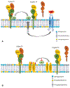
Mechanisms of protein disulphide isomerase (PDI)-mediated tissue factor (TF) decryption. (A) Cryptic TF contains free thiols. Cell activation by adenosine triphosphate (ATP) or other mediators results in the secretion of PDI and/or facilitates cell surface-associated PDI-dependent thiol-disulphide exchange reactions that lead to the formation of Cys186-Cys209 disulphide bond. The formation of Cys186-Cys209 disulphide bond results in a conformational change in TF–factor VIIa (FVIIa), favouring its interaction with factor X. (B) PDI, through its oxidoreductase activity, also modulates phosphatidylserine (PS) dynamics at the cell surface. Inhibition of PDI at the cell surface in endothelial cells was shown to inhibit the flippase and activate the floppase that results in the externalization of PS. PDI-dependent thiol-disulphide exchange reactions could also externalize PS. PDI-dependent thiol-disulphide exchange reactions that can switch on/off disulphide bonding in TF and modulate PS dynamics may closely cooperate in regulating TF pro-coagulant activity.
Fig. 4
Fig. 4
A coordinated mechanism of tissue factor (TF) activation. In resting cells, TF is maintained in an encrypted state by the presence of abundant sphingomyelin (SM) in the outer leaflet, sequestration of phosphatidylserine (PS) to the inner leaflet and TF with a reduced Cys186-Cys209 allosteric disulphide bond or a mixed disulphide bond. Adenosine triphosphate (ATP) activation of the P2×7 receptor (P2×7R) or other cell stimulus induces cell signalling that mobilizes acid-sphingomyelinase (ASM) from lysosomal compartment to the outer leaflet of the plasma membrane. Breakdown of SM by ASM releases TF from the SM-mediated encryption and alters membrane structure and fluidity leading to PS externalization and plasma membrane blebbing and shedding. ATP activation of the P2×7R also results in PS externalization and protein disulphide isomerase (PDI)-dependent thiol-disulphide exchange at the cell surface that results in the formation of Cys186-Cys209 disulphide bond in TF and alters TF conformation more favourable to the substrate binding. All events may work coordinately to fully activate TF at the cell surface and the release of TF+ MV.

References

    1. Rapaport SI, Rao LVM. The tissue factor pathway: how it has become a “prima ballerina”. Thromb Haemost 1995;74(01): 7–17 - PubMed
    1. Drake TA, Morrissey JH, Edgington TS. Selective cellular expression of tissue factor in human tissues. Implications for disorders of hemostasis and thrombosis. Am J Pathol 1989;134(05):1087–1097 - PMC - PubMed
    1. Fleck RA, Rao LVM, Rapaport SI, Varki N. Localization of human tissue factor antigen by immunostaining with monospecific, polyclonal anti-human tissue factor antibody. Thromb Res 1990;59(02):421–437 - PubMed
    1. Giesen PL, Rauch U, Bohrmann B, et al. Blood-borne tissue factor: another view of thrombosis. Proc Natl Acad Sci U S A 1999;96(05): 2311–2315 - PMC - PubMed
    1. Butenas S, Bouchard BA, Brummel-Ziedins KE, Parhami-Seren B, Mann KG. Tissue factor activity in whole blood. Blood 2005;105 (07):2764–2770 - PubMed