. 2019 Sep;235(3):555-568.
doi: 10.1111/joa.12971. Epub 2019 Mar 12.

Expression of ventral telencephalon transcription factors ASCL1 and DLX2 in the early fetal human cerebral cortex

Affiliations

Expression of ventral telencephalon transcription factors ASCL1 and DLX2 in the early fetal human cerebral cortex

Ayman Alzu'bi et al. J Anat. 2019 Sep.

Abstract

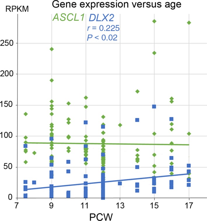
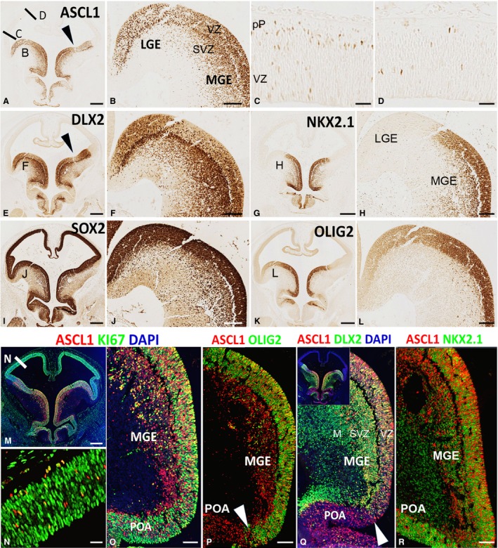
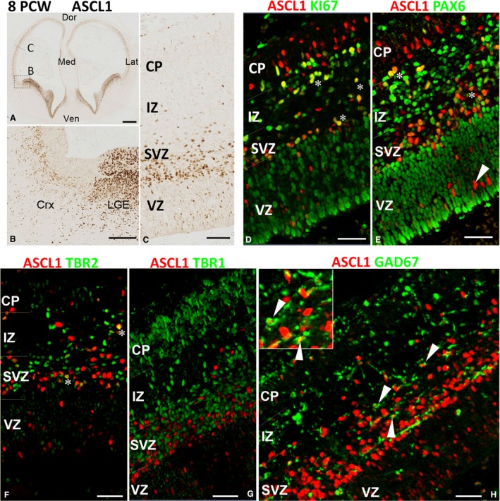
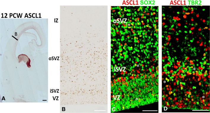
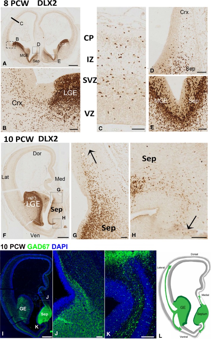
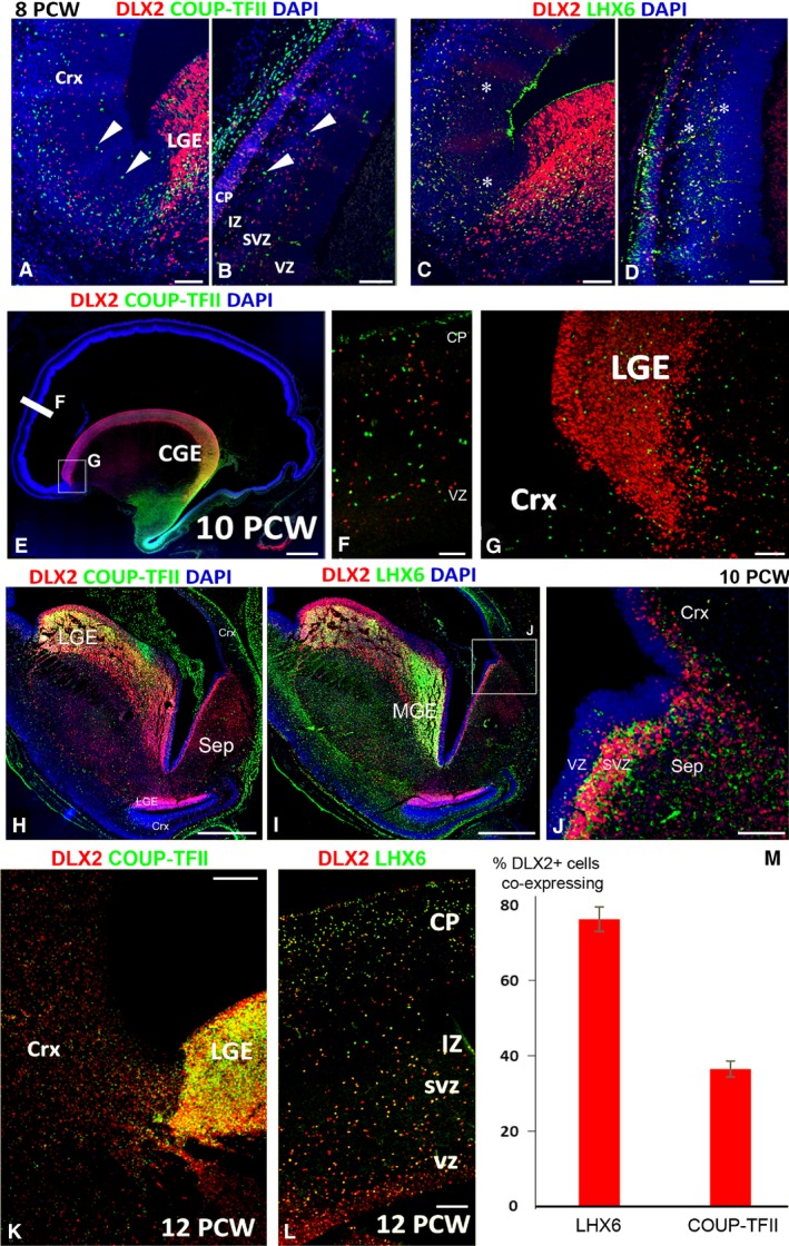
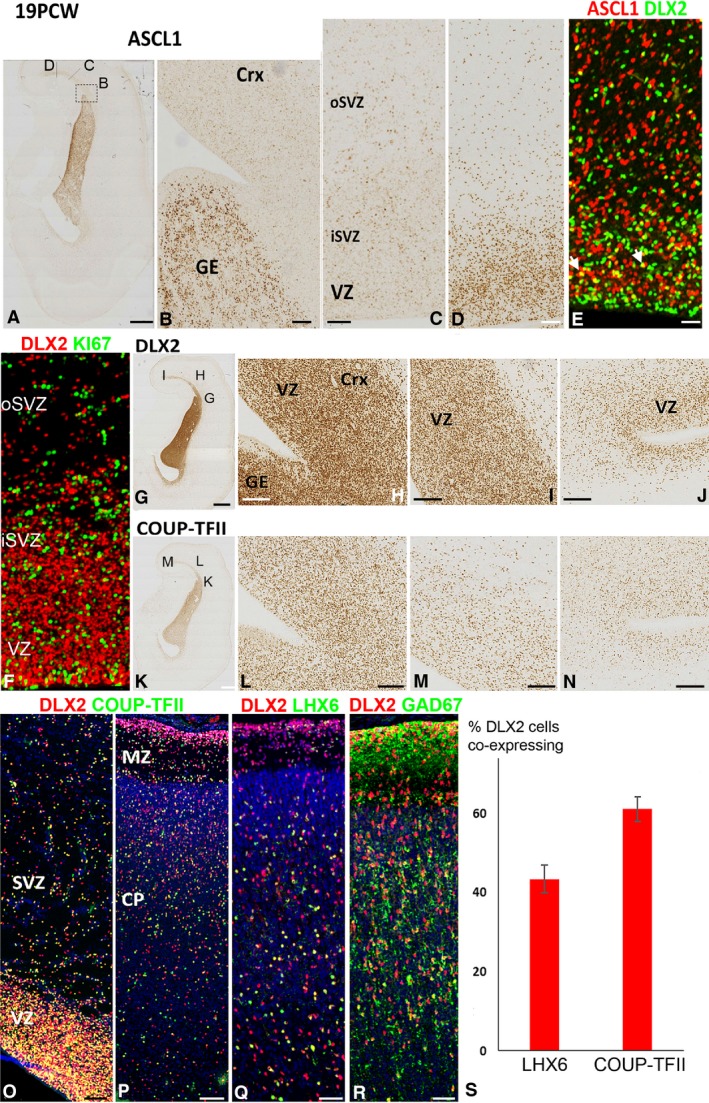
In rodent ventral telencephalon, diffusible morphogens induce expression of the proneural transcription factor ASCL1, which in turn induces expression of the transcription factor DLX2 that controls differentiation of cortical interneuron precursors and their tangential migration to the cerebral cortex. RNAseq analysis of human fetal samples of dorsal telencephalon revealed consistently high cortical expression of ASCL1 and increasing expression of DLX2 between 7.5 and 17 post-conceptional weeks (PCW). We explored whether cortical expression of these genes represented a population of intracortically derived interneuron precursors. Immunohistochemistry revealed an ASCL1+ /DLX2+ population of progenitor cells in the human ganglionic eminences between 6.5 and 12 PCW, but in the cortex there also existed a population of ASCL1+ /DLX2- progenitors in the subventricular zone (SVZ) that largely co-expressed cortical markers PAX6 or TBR2, although a few ASCL1+ /PAX6- progenitors were observed in the ventricular zone (VZ) and ASCL1+ cells expressing the interneuron marker GAD67 were present in the SVZ. Although rare in the VZ, DLX2+ cells progressively increased in number between 8 and 12 PCW across the cortical wall and the majority co-expressed LHX6 and originated either in the MGE, migrating to the lateral cortex, or from the septum, populating the medial wall. A minority co-expressed COUP-TFII, which identifies cells from the caudal ganglionic eminence (CGE). By 19 PCW, a significant increase in expression of DLX2 and ASCL1 was observed in the cortical VZ with a small proportion expressing both proteins. The DLX2+ cells did not co-express a cell division marker, so were not progenitors. The majority of DLX2+ cells throughout the cortical plate expressed COUP-TFII rather than LHX6+ . As the VZ declined as a proliferative zone it appeared to be re-defined as a migration pathway for COUP-TFII+ /DLX2+ interneurons from CGE to cortex. Therefore, in developing human cortex, ASCL1 expression predominantly marks a population of intermediate progenitors giving rise to glutamatergic neurons. DLX2 expression predominantly defines post-mitotic interneuron precursors.

Keywords: cortical inhibitory interneurons; development; dorsal telencephalon; ganglionic eminences.


Conflict of interest statement

The authors have no conflict of interest to declare.

Figures

Figure 1
Figure 1
Change in ASCL1 and DLX2 expression with age. RNAseq data from a range of cortical samples from anterior to posterior locations taken between 7.5 and 17 post‐conceptional weeks (PCW). ASCL1 showed high levels of expression maintained across the age range. DLX2 showed an increasing level of expression with age which was statistically significant (r = correlation coefficient; P = probability of correlation not being real). RPKM; normalised reads per kilobase of transcript per million mapped reads.
Figure 2
Figure 2
Expression of ASCL1, DLX2, and associated proteins at 6.5 PCW. (A) Shows high expression of ASCL1 in ganglionic eminences, preoptic area, and hypothalamus. Location of origin of panels (B,C,D) are marked. Arrowhead indicates boundary between LGE and MGE. (B) Shows ASCL1 expression was high in the ventricular zone (VZ) of both LGE and MGE and also high in the subventricular zone (SVZ) of the MGE. In the dorsal telencephalon expression was low, but higher laterally (C) than dorsally (D) with a few cells deep in the VZ but more present at the boundary of the VZ and the nascent post‐mitotic preplate (pP). (E) Shows high expression of DLX2 in ganglionic eminences, preoptic area, and hypothalamus with lower expression in the VZ of the ganglionic eminences and higher expression in the SVZ and overlying mantle. NKX2.1 expression was confined to MGE, preoptic area and parts of the hypothalamus (G,H). SOX2 expression marked all progenitor cells in the forebrain and illustrates the extent of the SVZ in the MGE (I,J) and strong expression of OLIG2 also delineated the MGE (K,L). Immunofluorescence double‐labelling showed that ASCL1+ cells of the telencephalon expressed cell division marker KI67 and thus were dividing neuroprogenitors (M–O). Co‐expression patterns of ASCL1 with OLIG2 (P) and DLX2 (Q) define the boundary between MGE and preoptic area (POA), although NKX2.1 is expressed in both regions (R). Scale bars: 500 μm (A,E,I,G,K,M), 100 μm (B,F,H,J,L,O,P,R for Q see P or R), 50 μm (C,D,N).
Figure 3
Figure 3
ASCL1 expression in the cortex at 8 PCW. (A) Anterior section of telencephalon; box shows the approximate location of panel (B) and the line of panel (C). Very high expression of ASCL1 in the ganglionic eminences and septum, and discernible expression in the cerebral cortex was observed (A,B). In the cortex, ASCL1 was predominantly expressed in subventricular zone (SVZ) and intermediate zone (IZ) with (D) many cells co‐expressing the marker for cell division KI67 (yellow, e.g. asterisks) and (E) the marker for radial glia, PAX6 (yellow, asterisks) in these locations. However, in the ventricular zone (VZ), co‐expression with either marker was rare (arrowhead). (F) ASCL1 was also co‐expressed with the marker of intermediate progenitor cells TBR2 (asterisks) but not with TBR1 (G) the marker for post‐mitotic neurons. (H) Co‐expression with GAD67, marker of GABAergic interneurons (arrowheads). Inset shows two examples of these double‐labelled cells at higher magnification. Dor, dorsal; Med, medial; Ven; ventral; Lat, lateral; CP; cortical plate. Scale bars: 500 μm (A), 100 μm (B), 50 μm (C–G) and 30 μm (H).
Figure 4
Figure 4
ASCL1 expression in the cortex at 12 PCW. (A) Section of telencephalon including MGE and LGE (high ASCL expression) with area B showing approximate location of panel (B). (B) Shows low expression of ASCL1 in the VZ but higher in the inner subventricular zone (iSVZ) and also throughout the outer subventricular zone (oSVZ). (C) Asterisks mark examples of double‐labelling of ASCL1 (red) with SOX2 (green, expressed by radial glia) and (D) mark examples of double‐labelling with TBR2 (expressed by intermediate progenitor cells). Scale bars:  1 mm (A), 100 μm (B) and 50 μm (C,D).
Figure 5
Figure 5
DLX2 expression 8–10 PCW. (A) DLX2 immunoreactivity in telencephalon at 8 PCW; bar and boxes show approximate location of panels (C,B,D). (B) Pallial/subpallial boundary. Dense expression of all layers of the lateral ganglionic eminence (LGE) with DLX2+ cells appearing to enter the cortex (Crx) principally through the SVZ. (C) Further dorsally in the cortical wall, DLX2+ cells are present in cortical plate (CP) intermediate zone (IZ) subventricular zone (SVZ) but with only a few in the ventricular zone (VZ). A few DLX2+ cells appear to migrate from the septum (Sep) to the dorsomedial cortex (D). Expression of DLX2 is continuous across the border between the medial ganglionic eminence (MGE) and septum, moderate in the ventricular layer and higher in the subventricular zone. (F) A more anterior section of the telencephalon at 10 PCW; high expression in the LGE, moderate in the septum. (G) Migration (arrow) of DLX2+ cells from septum to cortex dorsomedially. (H) Migration ventromedially. (I,J) Similar pattern of expression for GAD67, another marker of interneurons, that is highly expressed in the ganglionic eminences (GE) and septum (Sep), with GAD67+ cells appearing to migrate dorsally (J) and ventrally (K) from the septum. (L) A diagrammatic representation of the findings in (F–K) showing the lateral and medial pathways of migration of interneuron precursors from the subpallium to both dorsal and ventral cortex in the frontal lobe. Scale bars: 500 μm (A), 50 μm (B–E), 1 mm (F,I) and 100 μm (G,H,J,K).
Figure 6
Figure 6
Co‐expression of DLX2 with LHX6 and COUP‐TFII. (A) At 8 PCW, both DLX2+ cells (red) and COUP‐TFII+ cells (green) can be seen in the cortex (Crx) but the populations are non‐overlapping. Arrowheads show examples of COUP‐TFII+/DLX2 cells throughout the cortical wall (A,B). However, extensive co‐expression of DLX2 and COUP‐TFII (yellow, asterisks) was observed (C,D). The situation was similar at 10 PCW (E) with extensive co‐expression in the ventral caudal ganglionic eminence (CGE, yellow). No double‐labelled cells were present in the cortex or in the LGE (F,G). DLX2+/COUP‐TFII+ co‐expression (yellow) was observed in the LGE at the boundary with the lateral cortex but not at the LGE/ventral cortex boundary, or in the MGE or septum (H). However, DLX2 was co‐localised with LHX6 in the MGE (I) and also to a lesser extent in septum (J). By 12 PCW, the numbers of COUP‐TFII cells in the LGE had greatly increased and double‐labelled cells were present throughout the cortical wall (yellow, K,L). Nevertheless, cell counts demonstrated that a higher proportion of DLX2+ cells co‐expressed LHX6 than COUP‐TFII (M). LGE, lateral ganglionic eminence; CP, cortical plate; IZ, intermediate zone; SVZ, subventricular zone; VZ, ventricular zone. In (J) error bars represent the standard error of the mean. Scale bars: 100 μm (A–D,G,L), 1 mm (E), 50 μm (F) and 500 μm (H,I,K).
Figure 7
Figure 7
ASCL1 and DLX2 expression at 19 PCW. (A) Coronal section of cortex and ganglionic eminence immunostained for ASCL1 showing approximate location of panels (B,C,D). (B) High expression in the ganglionic eminence (GE) and very low expression in the lateral cortex which increases at more dorsomedial cortical locations (C,D) in both the inner and outer subventricular zone (iSVZ, oSVZ), but especially in the ventricular zone (VZ). (E) Co‐expression of ASCL1 (red) and DLX2 (green) was rare but observed in a few cells (yellow, e.g. arrowheads). (F) However, no co‐expression of DLX2 (red) with KI67 (green) was observed in the cortex. DLX2 expression was highest in the GE and in the lateral cortex close to the pallial/subpallial border, particularly in the VZ (G,H). More dorsomedially, the concentration of DLX2+ cells decreased (I,J). COUP‐TFII exhibited a similar pattern of expression (K–N), and DLX2 (red) and COUP‐TFII (green) were widely co‐expressed (yellow) throughout the cortical wall (O,P). A proportion of DLX2 cells also co‐expressed LHX6 (yellow, Q) and extensively co‐expressed GAD67 (R). At this stage of development, a higher proportion of DLX2+ cells co‐expressed COUP‐TFII than LHX6 (S). Scale bars: 2 mm (A,G,K), 200 μm (B), 25 μm (C–E for F see E) 200 μm (L,M,N) and 50 μm (O–R).

References

    1. Al‐Jaberi N, Lindsay S, Sarma S, et al. (2015) The early fetal development of human neocortical gabaergic interneurons. Cereb Cortex 25, 635–651. - PMC - PubMed
    1. Alzu'bi A, Lindsay SJ, Harkin LF, et al. (2017a) The transcription factors COUP‐TFI and COUP‐TFII have distinct roles in arealisation and GABAergic interneuron specification in the early human fetal telencephalon. Cereb Cortex 27, 4971–4987. - PMC - PubMed
    1. Alzu'bi A, Lindsay S, Kerwin J, et al. (2017b) Distinct cortical and sub‐cortical neurogenic domains for GABAergic interneuron precursor transcription factors NKX2.1, OLIG2 and COUP‐TFII in early fetal human telencephalon. Brain Struct Funct 222, 2309–2328. - PMC - PubMed
    1. Anders S, Pyl TP, Huber W (2014) HTSeq – A Python framework to work with high‐throughput sequencing data. Bioinformatics 32, 166–169. - PMC - PubMed
    1. Anderson SA, Eisenstat DD, Shi L, et al. (1997) Interneuron migration from basal forebrain to neocortex: dependence on Dlx genes. Science 278, 474–476. - PubMed

Publication types

MeSH terms

Substances