Infected chronic sinus secondary to a retained fragment of radial artery introducer sheath following percutaneous coronary intervention (PCI)
- PMID: 30862671
- PMCID: PMC6441256
- DOI: 10.1136/bcr-2018-227136
Infected chronic sinus secondary to a retained fragment of radial artery introducer sheath following percutaneous coronary intervention (PCI)
Abstract
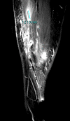
Coronary angiography and percutaneous coronary intervention (PCI) are frequently performed procedures in the UK and the developed world, with the radial artery becoming the preferred route of access. A chronically retained macroscopic fragment of radial artery introducer sheath is a very rare complication that has not, to our knowledge, been reported. We report the case of a 62-year-old woman who underwent PCI and developed a persisting infected sinus and abscess at the cannulation site despite multiple courses of antibiotics. Surgical exploration of the forearm recovered a foreign body that was found in the brachioradialis muscle and resembled a fragment of hydrophilic sheath. In conclusion, this case highlights that it is possible to leave macroscopic fragments of hydrophilic sheaths in situ. This is likely to be encountered during difficult access, especially during arterial spasm, and it is advised that the sheath and any other vascular access device is thoroughly inspected following removal.
Keywords: cardiovascular medicine; interventional cardiology; interventional radiology; orthopaedic and trauma surgery; orthopaedics.
© BMJ Publishing Group Limited 2019. No commercial re-use. See rights and permissions. Published by BMJ.
Conflict of interest statement
Competing interests: None declared.
Figures


References
-
- British Cardiovascular Intervention Society. National Audit of Percutaneous Coronary Interventions Annual Public Report. www.ucl.ac.uk/nicor/audits/adultpercutaneous/reports (accessed 19 Sep 2016).
Publication types
MeSH terms
LinkOut - more resources
Full Text Sources
Medical
Miscellaneous
