. 2019 Apr 26;294(17):7013-7024.
doi: 10.1074/jbc.RA119.007662. Epub 2019 Mar 12.

The retroviral accessory proteins S2, Nef, and glycoMA use similar mechanisms for antagonizing the host restriction factor SERINC5

Affiliations

The retroviral accessory proteins S2, Nef, and glycoMA use similar mechanisms for antagonizing the host restriction factor SERINC5

Iqbal Ahmad et al. J Biol Chem. .

Abstract

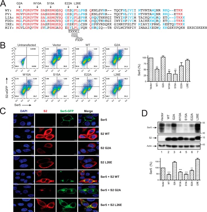
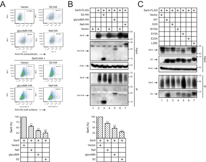
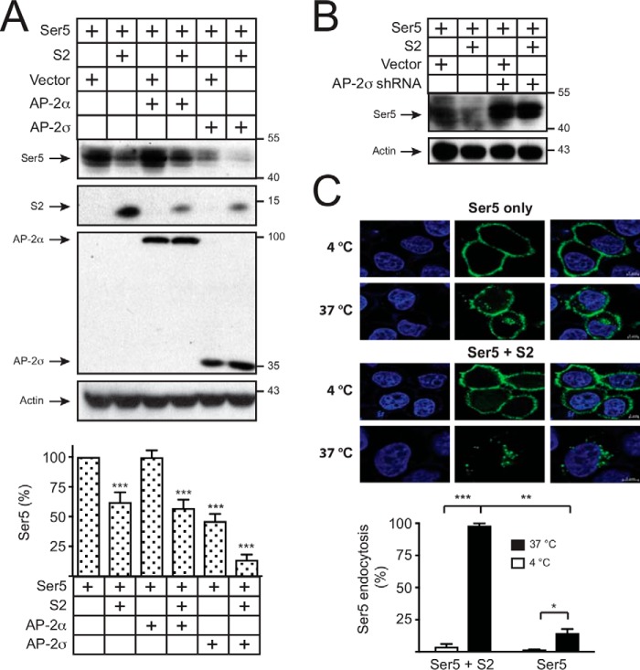
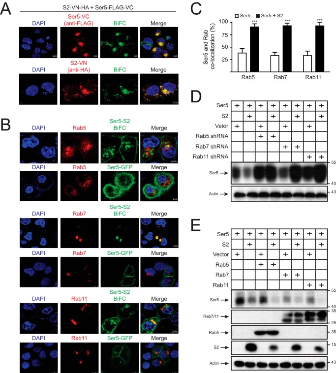
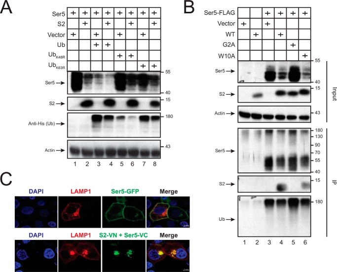
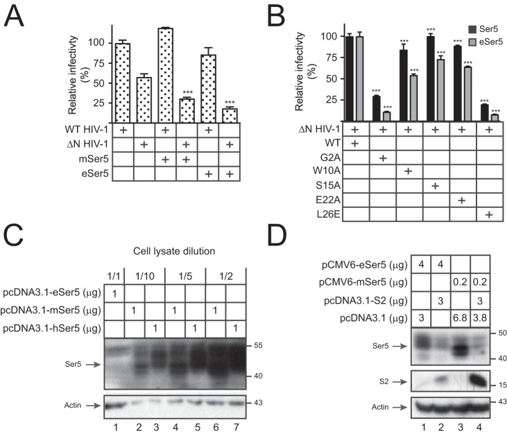
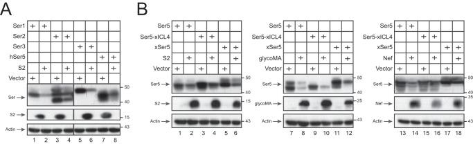
Serine incorporator 5 (SERINC5) is a recently identified restriction factor that blocks virus entry but is antagonized by three unrelated retroviral accessory proteins. The S2 protein from equine infectious anemia virus (EIAV) has been reported to reduce SERINC5 expression at steady-state levels likely via the endocytic pathway; however, the precise mechanism is still unclear. Here, we investigated how EIAV S2 protein down-regulates SERINC5 compared with down-regulation induced by Nef from HIV-1 and glycoMA proteins from murine leukemia virus (MLV). Using bimolecular fluorescence complementation (BiFC) assay and immunoprecipitation (IP), we detected an interaction between S2 and SERINC5. We found that this interaction relies on the S2 myristoylation site, indicating that it may occur on the plasma membrane. S2 internalized SERINC5 via receptor-mediated endocytosis and targeted it to endosomes and lysosomes, resulting in a ubiquitination-dependent decrease in SERINC5 expression at steady-state levels. Both BiFC and IP detected a glycoMA-SERINC5 interaction, but a Nef-SERINC5 interaction was detected only by BiFC. Moreover, S2 and glycoMA down-regulated SERINC5 more effectively than did Nef. We further show that unlike Nef, both S2 and glycoMA effectively down-regulate SERINC2 and also SERINC5 from Xenopus tropicalis (xSERINC5). Moreover, we detected expression of the equine SERINC5 (eSERINC5) protein and observed that its expression is much weaker than expression levels of SERINC5 from other species. Nonetheless, eSERINC5 had a strong antiviral activity that was effectively counteracted by S2. We conclude that HIV-1, EIAV, and MLV share a similar mechanism to antagonize viral restriction by host SERINC5.

Keywords: EIAV S2; HIV Nef; MLV glycoGag; SERINC5; host-pathogen interaction; innate immunity; restriction factor; viral protein; virus; virus entry.


Conflict of interest statement

The authors declare that they have no conflicts of interest with the contents of this article

Figures

Figure 1.
Figure 1.
S2 down-regulation of Ser5. A, WT and ΔN HIV-1 pseudoviruses were produced from 293T cells after transfection of 1 μg of Env expression vector pNLnΔBS and 1 μg of Env-deficient HIV-1 proviral vector pNLenCAT or pNLenCAT-Xh in the absence or presence of 1 μg of pBJ5-mSer5-FLAG and/or 2 μg of pcDNA3.1-S2-HA. Viruses were normalized by p24Gag ELISA, and viral infectivity was determined after infection of TZM-bI cells. The infectivity of WT viruses produced in the absence of Ser5 was set as 100%. B, 293T cells were transfected with 2 μg of pBJ5-mSer5-FLAG and 3 μg of pcDNA3.1-S2-HA or its empty vector. After 24 h, protein expression was detected by Western blotting. C, 293T cells were transfected with 0.1 μg of pCMV6-mSer5-FLAG and 2 μg of pcDNA3.1-S2-HA or its empty vector. After 24 h, cells were treated with MG132 (20 μm) or NH4Cl (20 μm) for 12 h, and the protein expression was analyzed by Western blotting. D, HeLa cells were transfected with 1 μg of pEGFP-N1-mSer5-FLAG and/or 2 μg of pcDNA3.1-S2-HA or pcDNA3.1-NefSF2-HA. The Ser5- and S2-cotransfected cells were treated with bafilomycin A1 (100 nm) for another 12 h. S2 and Nef proteins were stained with anti-HA followed by Alexa Fluor 647–conjugated goat anti-mouse and detected by confocal microscopy. E, HeLa cells were transfected with pEGFP-N1-mSer5-FLAG and pcDNA3.1-S2-HA or pcDNA3.1-NefSF2-HA. Cells were treated with bafilomycin A1 and stained with anti-HA as described (D). The frequency of S2/Ser5 and Nef/Ser5 double-positive cells was calculated by confocal microscopy, with that in treated cells set as 100%. Error bars in A, C, and E indicate S.E. from three independent experiments. **, p < 0.01.
Figure 2.
Figure 2.
Crucial S2 residues for Ser5 down-regulation. A, S2 protein sequences from six different EIAV strains are aligned, including AAC03764 (WY), U01888 (PV), AAK21109 (LIA), GU385359 (FDD), AFW99166 (IRE), and AFV61766 (MIY). Red, blue, and black represent residues completely, partially, or not conserved, and dashes indicate deletions. Targeted residues for mutagenesis including Gly2, Trp10, Ser15, Glu22, and Leu26 are indicated by arrowheads. The putative EXXXLL, YXXL, and PXXP motifs are also indicated. B, 293T cells were transfected with 2 μg of pBJ5-iFLAG-mSer5 and 3 μg of pEGFP-N1 vector expressing the indicated S2 proteins. Levels of Ser5 expression on the surface of EGFP-positive cells were analyzed by flow cytometry. Results are shown as relative values, with the value of Ser5 in the presence of the pEFGP-N1 vector set as 100%. C, HeLa cells were transfected with 1 μg of pEGFP-N1-mSer5-FLAG and 3 of μg pcDNA3.1 vector expressing the indicated S2 proteins. Doubly transfected cells were treated with bafilomycin A1. Ser5 and S2 localizations were detected by confocal microscopy as in Fig. 1D. D, 293T cells were transfected with 0.1 μg of pCMV6-mSer5-FLAG and 3 μg of pcDNA3.1 vector expressing the indicated S2 proteins. The Ser5 protein expression was detected by Western blotting. The relative Ser5 expression levels are shown by quantifying their intensity on Western blots, with the value of Ser5 in the presence of pcDNA3.1 vector set as 100%. Error bars in B and D indicate S.E. from three independent experiments. **, p < 0.01; ***, p < 0.001.
Figure 3.
Figure 3.
Detection of the S2–Ser5 interaction. A, 293T cells were transfected with 1 μg of pcDNA3.1-S2-HA, pcDNA3.1-glycoMA-HA, or pcDNA3.1-NefSF2-VN-HA alone or together with 100 ng of pBJ5-iHA-Ser5. Levels of S2, glycoMA, and Nef expression inside cells and levels of Ser5 expression on the cell surface were measured by flow cytometry. The relative Ser5 expression on the cell surface was calculated, with the value in the presence of pcDNA3.1 vector set as 100%. B, 293T cells were transfected with 0.5 μg of pCMV6-mSer5-FLAG and 12 μg of pcDNA3.1-S2-HA, pcDNA3.1-glycoMA-HA, or pcDNA3.1-NefSF2-HA. After immunoprecipitation by anti-FLAG, proteins in cell lysate (Input) and pulldown (IP) samples were analyzed by Western blotting. The relative Ser5 expression in cell lysate was calculated by quantifying the intensity on Western blots, with the value in the presence of the pcDNA3.1 vector set as 100%. C, 293T cells were transfected with 0.5 μg of pCMV6-mSer5-FLAG and 12 μg of pcDNA3.1 expressing WT or the indicated mutant S2 proteins. Proteins were pulled down and analyzed similarly. Error bars in A and B indicate S.E. from three independent experiments. ***, p < 0.001.
Figure 4.
Figure 4.
Detection of Ser5 endocytosis. A, 293T cells were transfected with 0.1 μg of pCMV6-mSer5-FLAG and 3 μg of pcDNA3.1-S2-HA in the presence of 1 μg of AP-2α or AP-2σ expression vector. AP-2α and AP-2σ were detected by anti-V5, Ser5 was detected by anti-FLAG, and S2 was detected by anti-HA. The relative Ser5 expression was calculated by quantifying the intensity on Western blots, with the value in the presence of pcDNA3.1 vector only set as 100%. B, 293T cells were transfected with 0.1 μg of pCMV6-mSer5-FLAG and 3 μg of pcDNA3.1-S2-HA in the presence of 4 μg of AP-2σ shRNA expression vector. Ser5 was detected by Western blotting. C, 293T cells were transfected with 1 μg of pBJ5-iFLAG-mSer5 and 3 μg of pcDNA3.1-S2-HA, and Ser5 endocytosis was detected at 4 and 37 °C. The levels of Ser5 endocytosis in the presence of S2 at 37 °C were set as 100%. Error bars in A and C indicate S.E. from three independent experiments. *, p < 0.05; **, p < 0.01; ***, p < 0.001.
Figure 5.
Figure 5.
Ser5 is targeted to endosomes. A, HeLa cells were transfected with 1 μg of pcDNA3.1-mSer5-FLAG-VC and 3 μg of pcDNA3.1-S2-VN-HA. After 24 h, cells were incubated with anti-FLAG or anti-HA followed by staining with Alexa Fluor 647–conjugated goat anti-mouse. The BiFC and immunofluorescence signals were detected by a confocal microscopy. B, HeLa cells were transfected with 1 μg of pCMV-mRFP-HA-Rab5a, pCMV-DsRed-2xHA-Rab7a, or pCMV-DsRed-2xHA-Rab11a in the presence of 1 μg of pEGFP-N1-mSer5-FLAG or 1 μg of pcDNA3.1-mSer5-FLAG-VC plus 3 μg of pcDNA3.1-S2-VN-HA. Fluorescence signals were detected by confocal microscopy. C, the colocalization of Ser5 with Rab small GTPases in B was statistically analyzed. Error bars indicate S.E. from three independent experiments. D and E, 293T cells were transfected with 0.1 μg of pCMV6-mSer5-FLAG and 3 μg of pcDNA3.1-S2-HA or its control vector in the presence of either 4 μg of Rab5, Rab7, and Rab11 shRNA expression vectors (D) or 1 μg of their expression vectors (E). Protein expressions were determined by Western blotting. ***, p < 0.001.
Figure 6.
Figure 6.
Ser5 is targeted to lysosomes. A, 293T cells were transfected with 0.1 μg of pCMV6-mSer5-FLAG and 3 μg of pcDNA3.1-S2-HA or its control vector in the presence of 1 μg of WT Ub or its mutant (UbK48R or UbK63R) expression vectors. Protein expressions were detected by Western blotting. B, Ser5 and the indicated S2 proteins were expressed, and proteins were pulled down and analyzed as in Fig. 3B. C, HeLa cells were transfected with 1 μg of pCMV-LAMP1-mRFP in the presence of 1 μg of pEGFP-N1-mSer5-FLAG or 1 μg of pcDNA3.1-mSer5-VN-HA plus 3 μg of pcDNA3.1-S2-FLAG-VC. Fluorescence signals were detected by confocal microscopy.
Figure 7.
Figure 7.
Analysis of eSer5 antiviral activity and sensitivity to S2. A, WT and ΔN HIV-1 pseudoviruses were produced from 293T cells in the absence or presence of 0.2 μg of pcDNA3.1-mSer5-FLAG or pcDNA3.1-eSer5-FLAG. Viral infectivity was measured and presented as in Fig. 1A. B, ΔN HIV-1 pseudoviruses were produced from 293T cells in the presence of pcDNA3.1-mSer5-FLAG or pcDNA3.1-eSer5-FLAG and the indicated S2 expression vectors. Viral infectivity was measured and presented as in Fig. 1A. C, 293T cells were transfected with 1 μg of pcDNA3.1-eSer5-FLAG, pcDNA3.1-mSer5-FLAG, or pcDNA3.1-hSer5-FLAG. Lysate from cells expressing human and murine Ser5 was diluted as indicated, and Ser5 expressions were compared by Western blotting. D, 293T cells were transfected with 3 μg of pcDNA3.1-S2-HA and 4 μg of pCMV6-eSer5-FLAG or 0.2 μg of pCMV6-mSer5-FLAG. Ser5 and S2 expressions were determined by Western blotting. Error bars in A and B indicate S.E. from three independent experiments. ***, p < 0.001.
Figure 8.
Figure 8.
Broadness of the S2 antagonism. A, 293T cells were transfected with 0.2 μg of pCMV6-mSer1-FLAG, pCMV6-mSer2-FLAG, pCMV6-mSer3-FLAG, or pCMV6-hSer5-FLAG in the presence of 3 μg of pcDNA3.1-S2-HA or its control vector. SERINC and S2 expressions were determined by Western blotting. B, 293T cells were transfected with 3 μg of pcDNA3.1-S2-HA, pcDNA3.1-glycoMA-HA, pcDNA3.1-NefSF2-HA, or its control vector and 0.2 μg of pCMV6-mSer5-FLAG, pCMV6-Ser5-xICL4-FLAG, or pCMV6-xSer5-FLAG, and their expressions were detected by Western blotting.

References

    1. Inuzuka M., Hayakawa M., and Ingi T. (2005) Serinc, an activity-regulated protein family, incorporates serine into membrane lipid synthesis. J. Biol. Chem. 280, 35776–35783 10.1074/jbc.M505712200 - DOI - PubMed
    1. Rosa A., Chande A., Ziglio S., De Sanctis V., Bertorelli R., Goh S. L., McCauley S. M., Nowosielska A., Antonarakis S. E., Luban J., Santoni F. A., and Pizzato M. (2015) HIV-1 Nef promotes infection by excluding SERINC5 from virion incorporation. Nature 526, 212–217 10.1038/nature15399 - DOI - PMC - PubMed
    1. Usami Y., Wu Y., and Göttlinger H. G. (2015) SERINC3 and SERINC5 restrict HIV-1 infectivity and are counteracted by Nef. Nature 526, 218–223 10.1038/nature15400 - DOI - PMC - PubMed
    1. Zhang X., Zhou T., Yang J., Lin Y., Shi J., Zhang X., Frabutt D. A., Zeng X., Li S., Venta P. J., and Zheng Y. H. (2017) Identification of SERINC5-001 as the predominant spliced isoform for HIV-1 restriction. J. Virol. 91, e00137–17 10.1128/JVI.00137-17 - DOI - PMC - PubMed
    1. Sood C., Marin M., Chande A., Pizzato M., and Melikyan G. B. (2017) SERINC5 protein inhibits HIV-1 fusion pore formation by promoting functional inactivation of envelope glycoproteins. J. Biol. Chem. 292, 6014–6026 10.1074/jbc.M117.777714 - DOI - PMC - PubMed

Publication types

Substances

LinkOut - more resources