. 2019 May 3;294(18):7388-7402.
doi: 10.1074/jbc.RA119.007656. Epub 2019 Mar 12.

Phosphorylated tyrosine 93 of hepatitis C virus nonstructural protein 5A is essential for interaction with host c-Src and efficient viral replication

Affiliations

Phosphorylated tyrosine 93 of hepatitis C virus nonstructural protein 5A is essential for interaction with host c-Src and efficient viral replication

Stefan Klinker et al. J Biol Chem. .

Abstract

The hepatitis C virus (HCV) nonstructural protein 5A (NS5A) plays a key role in viral replication and virion assembly, and the regulation of the assembly process critically depends on phosphorylation of both serine and threonine residues in NS5A. We previously identified SRC proto-oncogene, nonreceptor tyrosine kinase (c-Src), as an essential host component of the HCV replication complex consisting of NS5A, the RNA-dependent RNA polymerase NS5B, and c-Src. Pulldown assays revealed an interaction between NS5A and the Src homology 2 (SH2) domain of c-Src; however, the precise binding mode remains undefined. In this study, using a variety of biochemical and biophysical techniques, along with molecular dynamics simulations, we demonstrate that the interaction between NS5A and the c-Src SH2 domain strictly depends on an intact phosphotyrosine-binding competent SH2 domain and on tyrosine phosphorylation within NS5A. Detailed analysis of c-Src SH2 domain binding to a panel of phosphorylation-deficient NS5A variants revealed that phosphorylation of Tyr-93 located within domain 1 of NS5A, but not of any other tyrosine residue, is crucial for complex formation. In line with these findings, effective replication of subgenomic HCV replicons as well as production of infectious virus particles in mammalian cell culture models were clearly dependent on the presence of tyrosine at position 93 of NS5A. These findings indicate that phosphorylated Tyr-93 in NS5A plays an important role during viral replication by facilitating NS5A's interaction with the SH2 domain of c-Src.

Keywords: NS5A; Src; Src homology 2 domain (SH2 domain); hepatitis C virus (HCV); liver disease; nonstructural protein 5A; phosphotyrosine; viral replication; virion assembly.


Conflict of interest statement

The authors declare that they have no conflicts of interest with the contents of this article

Figures

Figure 1.
Figure 1.
Pulldown analysis of NS5A with WT and pTyr-binding–deficient SH2 domains. Huh-9-13 hepatocyte lysates were subjected to pulldown experiments with recombinantly expressed and purified SH2 domain-containing constructs as indicated. NS5A was visualized by immunoblotting. Controls were performed with inactivated beads and immobilized GST. NS5A binding to NHS beads and to GST was negligible. Huh-9-13 total protein (1 and 2.5 μg) was used as the positive control.
Figure 2.
Figure 2.
Schematic representation of Tyr-containing peptides derived from NS5A (A) and screening of NS5A tyrosine residues for binding to c-Src–SH2 (B). Binding of the GST–c-Src constructs to the various immobilized biotinylated peptides was measured colorimetrically. Binding was normalized to the best binder (pTyr-93), which was arbitrarily set to 100. Mean and S.D. of n = 3 experiments are shown.
Figure 3.
Figure 3.
Binding affinities of NS5A-derived peptides to c-Src–derived constructs determined by fluorescence polarization. Dissociation constants (Kd) of different NS5A peptides toward c-Src–ΔSH1 (A), c-Src–SH2 (B), and c-Src–SH3SH2 (C) are plotted on a logarithmic scale. Raw data and data fits are shown in Fig. S2.
Figure 4.
Figure 4.
Analysis of NS5A–D2D3ELKpY binding to c-Src–ΔSH1 via BLI. Immobilized NS5A–D2D3ELKpY was incubated with c-Src–ΔSH1 at a wide range of concentrations (A). Kd was determined to be 18 ± 4 μm (B).
Figure 5.
Figure 5.
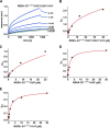
Interaction of phosphorylated and nonphosphorylated NS5A–D1 with WT or pTyr-binding–deficient c-Src–ΔSH1. BLI sensorgrams of analyte NS5A–D1ELKpY binding to the c-Src–ΔSH1 ligand (A) yield a Kd of 0.47 ± 0.1 μm (B). C, c-Src–ΔSH1 ligand was incubated with nonphosphorylated NS5A–D1. No binding was observed up to 40 μm analyte. D, c-Src–ΔSH1 R173K ligand was incubated with NS5A–D1ELKpY. No binding was observed up to 12 μm analyte.
Figure 6.
Figure 6.
Interaction of NS5A–D1 mutants with immobilized c-Src–ΔSH1 revealing the critical pTyr for SH2 binding. BLI sensorgrams and fits for different NS5A–D1 variants (analyte) and c-Src–ΔSH1 (ligand) are shown. A, c-Src–ΔSH1 was incubated with NS5A–D1ELKpY Y93F/Y129F/Y161F. Plotting of yinf against NS5A–D1ELKpY Y93F/Y129F/Y161F concentration (B) suggests the Kd to be above 40 μm. The remaining panels illustrate c-Src–ΔSH1 interaction with NS5A–D1ELKpY Y93F (C), NS5A–D1ELKpY Y129F (D), and NS5A–D1ELKpY Y161F (E), respectively. Kd values were determined as 1.5 ± 0.5 μm for NS5A–D1ELKpY Y129F and 2.1 ± 0.8 μm for NS5A–D1ELKpY Y161F. No binding of NS5A–D1ELKpY Y93F was observed in the concentration range applied. The raw data sensorgrams and fits are shown in Fig. S3.
Figure 7.
Figure 7.
Analysis of c-Src–ΔSH1 binding to immobilized NS5A–D1ELKpY Y129F/Y161F, confirming that pTyr-93 is critical for SH2 binding. BLI measurements were performed using the NS5A–D1ELKpY Y129F/Y161F mutant as ligand and c-Src–ΔSH1 as analyte. Sensorgrams (A) and fit (B) yield a Kd of 0.7 ± 0.2 μm.
Figure 8.
Figure 8.
Substitution of Tyr-93 with phenylalanine in NS5A severely impairs HCV replication and virus production in human hepatoma cells. A, Huh-7 cells were transfected with either WT HCV replicon plasmid pFK-I377/NS3-NS3′ (Rep) or the mutated plasmid pFK-I377/NS3-NS3′-Y93F (Rep Y93F). B and C, Huh-7.5 cells were transfected with either WT HCVcc JC1 plasmid pFK-JFH1J6C-846_dg (JC1) or the mutated plasmid pFK-JFH1J6C-846_dg-Y93F (JC1 Y93F). A and C, total mRNA was determined by semi-quantitative rtPCR, and results were calculated using the ΔΔCT method and SDHA as the control gene. Data are provided in relation to the respective control cells (Rep or JC1, set as 100) and are depicted as means ± S.D. of at least three independent experiments. The respective differences were significant (p = 0.037 and p = 0.032, respectively) as indicated by the one-tailed Mann-Whitney test. B, virus was collected from the supernatants, concentrated by PEG precipitation and infective titers determined by the TCID50 assay. Significant differences (p = 0.029) were confirmed using the one-tailed Mann-Whitney test.
Figure 9.
Figure 9.
Mechanistic model illustrating formation of the HCV replication complex, with a focus on the role of tyrosine phosphorylation events. A, in its basal state, the kinase domain of c-Src is kept in a restrained, inactive conformation by intramolecular interactions of the regulatory SH2 and SH3 domains, which bind to pTyr-530 close to the C terminus and to a proline-rich motif located in the SH2-kinase linker, respectively (47). B, upon NS5A phosphorylation at residue Tyr-93, NS5A–D1 displays a canonical, high-affinity binding site for c-Src–SH2; as a result, c-Src is recruited to the replication complex in its activated form, and HCV replication can occur. Other low-affinity interactions (21, 28) between LCS II or D2 of NS5A and the SH3 domain may also contribute (black dotted arrows). NS5B is complexed through c-Src–SH3 (gray dotted arrow (25) and NS5A (black broken arrows (35) interactions. For the sake of clarity, the dimerization site of NS5A is indicated without displaying a full dimer situation (refer to Fig. S6 for a discussion of steric restraints). DAAs like daclatasvir can bind to NS5A lacking phosphorylated Tyr-93 but are ineffective in the presence of pTyr-93. Regulatory tyrosine phosphorylation in c-Src is highlighted in yellow; NS5A–D1 phosphorylation at Tyr-93 is marked in red (abbreviations used are: AH, amphipathic helix; TD, transmembrane domain).

References

    1. Bode J. G., Brenndörfer E. D., Karthe J., and Häussinger D. (2009) Interplay between host cell and hepatitis C virus in regulating viral replication. Biol. Chem. 390, 1013–1032 - PubMed
    1. Moradpour D., Penin F., and Rice C. M. (2007) Replication of hepatitis C virus. Nat. Rev. Microbiol. 5, 453–463 10.1038/nrmicro1645 - DOI - PubMed
    1. Paul D., Hoppe S., Saher G., Krijnse-Locker J., and Bartenschlager R. (2013) Morphological and biochemical characterization of the membranous hepatitis C virus replication compartment. J. Virol. 87, 10612–10627 10.1128/JVI.01370-13 - DOI - PMC - PubMed
    1. Romero-Brey I., Berger C., Kallis S., Kolovou A., Paul D., Lohmann V., and Bartenschlager R. (2015) NS5A domain 1 and polyprotein cleavage kinetics are critical for induction of double-membrane vesicles associated with hepatitis C virus replication. MBio 6, e00759 - PMC - PubMed
    1. Tellinghuisen T. L., Marcotrigiano J., Gorbalenya A. E., and Rice C. M. (2004) The NS5A protein of hepatitis C virus is a zinc metalloprotein. J. Biol. Chem. 279, 48576–48587 10.1074/jbc.M407787200 - DOI - PubMed

Publication types

LinkOut - more resources