. 2019 Mar 12;9(1):4227.
doi: 10.1038/s41598-019-40588-5.

Surveillance of cell wall diffusion barrier integrity modulates water and solute transport in plants

Affiliations

Surveillance of cell wall diffusion barrier integrity modulates water and solute transport in plants

Peng Wang et al. Sci Rep. .

Abstract

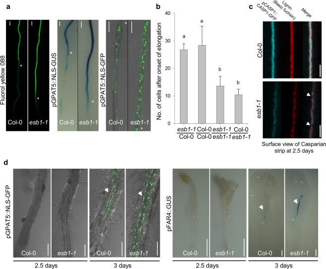
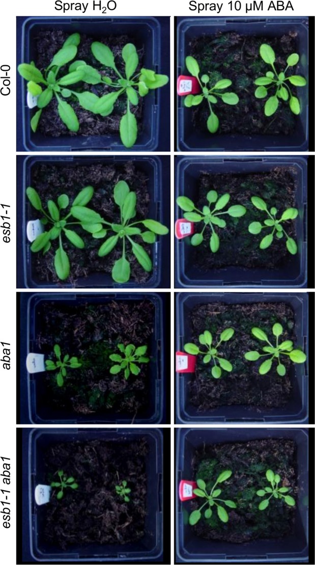
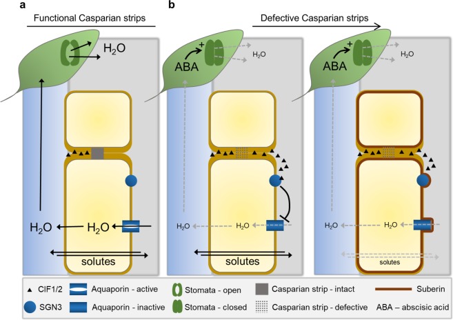
The endodermis is a key cell layer in plant roots that contributes to the controlled uptake of water and mineral nutrients into plants. In order to provide such functionality the endodermal cell wall has specific chemical modifications consisting of lignin bands (Casparian strips) that encircle each cell, and deposition of a waxy-like substance (suberin) between the wall and the plasma membrane. These two extracellular deposits provide control of diffusion enabling the endodermis to direct the movement of water and solutes into and out of the vascular system in roots. Loss of integrity of the Casparian strip-based apoplastic barrier is sensed by the leakage of a small peptide from the stele into the cortex. Here, we report that such sensing of barrier integrity leads to the rebalancing of water and mineral nutrient uptake, compensating for breakage of Casparian strips. This rebalancing involves both a reduction in root hydraulic conductivity driven by deactivation of aquaporins, and downstream limitation of ion leakage through deposition of suberin. These responses in the root are also coupled to a reduction in water demand in the shoot mediated by ABA-dependent stomatal closure.


Conflict of interest statement

The authors declare no competing interests.

Figures

Figure 1
Figure 1
Enhanced endodermal suberin in esb1-1 is driven by loss-of-function of ESB1 in roots. (a) Suberin staining with Fluorol yellow 088, promoter activity of suberin biosynthetic gene GPAT5 revealed with GUS staining and nuclear localized GFP (NLS-GFP), (bottom part: toward the root tip; top part: toward the hypocotyl) in 7 day-old seedlings. The white asterisks indicate the beginning of continuous Fluorol yellow 088 staining, GUS expression or GFP in either Col-0 or esb1-1. Scale bars: 100 μm. (b) Number of endodermal cells from the first fully expanded cell to the beginning of continuous Fluorol Yellow 088 staining in the lateral roots of 2 week-old grafted plants, including esb1-1 shoot/wild-type Col-0 root grafted, self-grafted wild-type Col-0, self-grafted esb1-1, and Col-0 shoot/esb1-1 root grafted. Different letters indicate the significant differences between means in pairwise comparison by Duncan’s MRT test (α = 0.05), means ± SE, n = 7. Fluorescence of lignin stained with basic fuchsin and pCASP1::CASP1-GFP (c) in wild-type Col-0 and esb1-1 in 2.5 day-old seedlings. The white arrows in (c) represent the disrupted lignification/CASP1-GFP localisation in esb1-1 compared to wild-type Col-0. Scale bars: 5 μm. GFP fluorescence and GUS staining (d) in suberin biosynthetic gene promoter reporter lines pGPAT5::NLS-GFP and pFAR4::GUS, respectively, at 2.5 days and 3 days after sowing in wild-type Col-0 and esb1-1. The white arrowheads show GFP fluorescence and GUS activity in esb1-1 in 3 day-old plants. Scale bars (d, left panel): 100 μm, (d, right panel): 1 mm, n = 3 roots were observed each time point and genotype, and representative pictures shown.
Figure 2
Figure 2
Whole root solute leakage, hydraulic conductivity and apoplastic diffusion barriers. (a) Solute leakage into the xylem in 21 day-old plants grown hydroponically subjected to increasing NaCl concentrations. The selectivity of the root to NaCl (σNaCl) was calculated as described in Method section (ANOVA Tukey’s HSD, means ± SE, n = 15–18, pooled from 3 biological replicates, p < 0.05). (b) Total root hydraulic conductivity (Lpr) of 21 day-old plants grown hydroponically (ANOVA Tukey’s HSD, means ± SE, n = 18–22, pooled from 3 biological replicates, p < 0.05). (c) Effect of the CIF2 peptide and the absence of its receptor (sgn3-3 mutant) on total root hydraulic conductivity of 21 day-old plants grown hydroponically (ANOVA Tukey’s HSD, means ± SE, n = 8–16, pooled from 3 biological replicates, p < 0.05). (d) Effect of the loss of function of the aquaporins PIP2;1 and PIP2;2 in the pip2;1 pip2;2 double mutant on endodermal suberisation. 5 day-old seedlings were stained with Fluorol Yellow 088. Quantification (%) of suberization along the root axis, distinguishing three differentiation stages, non-suberized, patchy and continuous (bottom panel) are presented (t-test, means ± SD, n = 4, p < 0.05). (e) Expression pattern of the suberisation reporter line pGPAT5::mCITRINE-SYP122. 5 day-old seedlings were treated with 0 and 10 µM sodium azide (NaN3) for 6 h. mCITRINE-SYP122 expression was quantified along the root axis, distinguishing three differentiation stages: continuous, patchy and no expression. Values are expressed as a percentage of root length (t-test, means ± SD, n = 6, p < 0.05).
Figure 3
Figure 3
Activation of ABA signalling at the endodermis is not involved in the enhanced deposition of suberin. Fluorol Yellow 088 staining for suberin in 5 day-old WT, pELTP::abi1-1, esb1-1 and pELTP::abi1-1esb1-1 roots. Pictures taken in similar parts of the root (upper panels) and quantification (%) of suberization along the root axis, distinguishing three differentiation stages, non-suberized, patchy and continuous (bottom panel) are presented (means ± SD, n ≥ 10). Scale bar, 50 μm.
Figure 4
Figure 4
ABA biosynthesis is essential for physiological compensation of the esb1 defect. Phenotype of Col-0, esb1-1, aba1 and esb1-1aba1 plants grown 26 days in the greenhouse at the early reproductive stage. The plants were sprayed with water or with 10 µM ABA.
Figure 5
Figure 5
Model summarising the integration between apoplastic endodermal diffusion barriers, hydraulic conductivity, solute permeability and stomatal conductance. (a) Functioning Casparian strips at the endodermis prevent the apoplastically localised peptide CIF1&2 from diffusing from the stele, across the endodermis, and into the cortex. (b) Defective Casparian strips are detected by leakage of CIF1&2 into the cortical apoplast where the peptides are sensed by binding to SGN3, signalling inactivation of aquaporins. This leads to reduced hydraulic conductivity and closure of stomates in leaves through a process mediated locally by ABA. Inactivation of aquaporins leads to the enhanced deposition of suberin which reduces solute conductivity across the root into and out of the xylem.

References

    1. Geldner N. The endodermis. Annu. Rev. Plant Biol. 2013;64:531–558. doi: 10.1146/annurev-arplant-050312-120050. - DOI - PubMed
    1. Andersen TG, Barberon M, Geldner N. Suberization — the second life of an endodermal cell. Current Opinion in Plant Biology. 2015;28:9–15. doi: 10.1016/j.pbi.2015.08.004. - DOI - PubMed
    1. Hosmani PS, et al. Dirigent domain-containing protein is part of the machinery required for formation of the lignin-based Casparian strip in the root. Proc. Natl. Acad. Sci. USA. 2013;110:14498–14503. doi: 10.1073/pnas.1308412110. - DOI - PMC - PubMed
    1. Pfister, A. et al. A receptor-like kinase mutant with absent endodermal diffusion barrier displays selective nutrient homeostasis defects. Elife3 (2014). - PMC - PubMed
    1. Kamiya, T. et al. The MYB36 transcription factor orchestrates Casparian strip formation. Proc. Natl. Acad. Sci. USA 201507691, 10.1073/pnas.1507691112 (2015). - PMC - PubMed

Publication types