Review
. 2019 Feb 22:12:1497-1508.
doi: 10.2147/OTT.S174316. eCollection 2019.

The surgical perspective in precision treatment of diffuse gliomas

Affiliations
Review

The surgical perspective in precision treatment of diffuse gliomas

Niklas Thon et al. Onco Targets Ther. .

Abstract

Over the last decade, advances in molecular and imaging-based biomarkers have induced a more versatile diagnostic classification and prognostic evaluation of glioma patients. This, in combination with a growing therapeutic armamentarium, enables increasingly individualized, risk-benefit-optimized treatment strategies. This path to precision medicine in glioma patients requires surgical procedures to be reassessed within multidimensional management considerations. This article attempts to integrate the surgical intervention into a dynamic network of versatile diagnostic characterization, prognostic assessment, and multimodal treatment options in the light of the latest 2016 World Health Organization (WHO) classification of diffuse brain tumors, WHO grade II, III, and IV. Special focus is set on surgical aspects such as resectability, extent of resection, and targeted surgical strategies including minimal invasive stereotactic biopsy procedures, convection enhanced delivery, and photodynamic therapy. Moreover, the influence of recent advances in radiomics/radiogenimics on the process of surgical decision-making will be touched.

Keywords: biomarker; cytoreductive surgery; extent of resection; metabolic imaging; molecular markers; personalized medicine; precision medicine; prognosis; stereotactic biopsy.


Conflict of interest statement

Disclosure The authors report no conflicts of interest in this work.

Figures
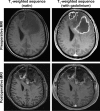
Figure 1
Figure 1
Tumor debulking. 64-year-old male patient with clinical deterioration (headache, motoric aphasia, cognitive impairment) due to a large, bifrontal space-occupying contrast-enhancing glioblastoma WHO grade IV, IDH wild-type. Upfront biopsy revealed a glioblastoma WHO grade IV, IDH-wildtype, with an unmethylated MGMT promoter status. Maximal safe resection was performed in order to relieve burden from space occupying effect and to stabilize the patient before chemoradiation could be initiated. Early postoperative T1-weighted sequences (with and without gadolinium) confirmed an extensive bifrontal tumor debulking with some hemorrhagic imbibition of the resection cavity. Clinically, the patient stabilized and the aphasia completely resolved. Chemoradiation could be initiated two weeks after surgery. Abbreviations: IDH, isocitrate dehydrogenasis; MGMT, O6-methylguanine-DNA-methyltransferase; MRI, magnetic resonance imaging; WHO, World Health Organization.
Figure 2
Figure 2
Volumetric assessment of EOR. Example of 60-year-old patient suffering from a large, highly vascularized, space-occupying IDH-wildtype, MGMT unmethylated glioblastoma of the right mesio-temporal lobe with critical involvement of the basal ganglia and compression of the midbrain. A subtotal resection of the almost spherical tumor formation (left side, highlighted in orange) has been achieved without perioperative morbidity. The postoperative control MRI confirmed an extensive resection with sufficient decompression of vulnerable structures and some minor complex shaped, multifocal tumor remnants (yellow). The respective volumes were calculated using a semiautomatically three-dimensional calculation tool (SmartBrush®, Brainlab, Feldkirchen, Germany) indicating an EOR of 91.4%. Abbreviations: ceMRA, contrast-enhanced magnetic imaging angiography; EOR, extent of resection; IDH, isocitrate dehydrogenasis; MGMT, O6-methylguanine-DNA-methyltransferase; MRI, magnetic resonance imaging.
Figure 3
Figure 3
Personalized surgical strategy. (A) 42-year-old male patient with a newly diagnosed tumor of the left frontal lobe. (i) Representative T2-weighted axial magnetic resonance imaging sequence at first diagnosis. (ii) Representative axial (top) and sagittal (below) reconstructions in F-FET PET metabolic imaging. (iii) Dynamic analysis in F-FET PET indicating exclusively increasing time activity curves (each line represents a measurement of dynamic F-FET uptake within one voxel over time) within the tumor tissue indicating a WHO grade II glioma. Histopathological evaluation of targeted tissue samples (by stereotactic biopsy) confirmed an IDH mutant, 1p/19q co-deleted oligodendroglioma WHO grade II. Neoadjuvant temozolomid chemotherapy was initiated by the interdisciplinary tumor board. At this time, any surgical risk as well as upfront irradiation were unacceptable for the busy managing director. (B) Follow-up after 6 cycles of temozolomide 5/28 protocol did not confirm any improvement with respect to the size of the tumor formation as being shown in (i) a representative axial T2-weighted sequence. (ii) Also, a stable tumor formation was confirmed by follow-up F-FET PET. Thereafter, maximal safe resection was recommended by the interdisciplinary tumor board. (C) Representative postoperative axial T2-weighted sequence. No new neurological deficit was seen. Thereafter, adjuvant irradiation was withheld due to stable tumor formation until last follow-up MRI. Abbreviations: F-FET, O-(2-[(18)F]-fluoroethyl)-L-tyrosine; IDH, isocitrate dehydrogenase; MRI, magnetic resonance imaging; PET, positron emission tomography; WHO, World Health Organization.

References

    1. Ostrom QT, Gittleman H, Liao P, et al. CBTRUS statistical report: primary brain and other central nervous system tumors diagnosed in the United States in 2010–2014. Neuro Oncol. 2017;19(suppl_5):v1–v88. - PMC - PubMed
    1. Louis DN, Perry A, Reifenberger G, et al. The 2016 World Health organization classification of tumors of the central nervous system: a summary. Acta Neuropathol. 2016;131(6):803–820. - PubMed
    1. Dunn GP, Andronesi OC, Cahill DP. From genomics to the clinic: biological and translational insights of mutant IDH1/2 in glioma. Neurosurg Focus. 2013;481(2):E2. - PubMed
    1. Weller M, van den Bent M, Tonn JC, et al. European association for neuro-oncology (EANO) guideline on the diagnosis and treatment of adult astrocytic and oligodendroglial gliomas. Lancet Oncol. 2017;18(6):e315–e329. - PubMed
    1. Ringel F, Pape H, Sabel M, et al. Clinical benefit from resection of recurrent glioblastomas: results of a multicenter study including 503 patients with recurrent glioblastomas undergoing surgical resection. Neuro Oncol. 2016;18(1):96–104. - PMC - PubMed