

Free-Flap Reconstruction of the Mandible
- PMID: 30863212
- PMCID: PMC6408245
- DOI: 10.1055/s-0039-1677791
Free-Flap Reconstruction of the Mandible
Abstract
Mandible reconstruction has evolved over the years with advances in surgical options and three-dimensional technology. Although nonvascularized bone grafting is still used, vascularized flaps show advantages with immediate reconstruction, the possibility of immediate dental implants, and the ability to reconstruct composite defects of both soft tissue and bone. This article discusses current vascularized techniques for mandible reconstruction. While each reconstructive method has advantages and disadvantages, a defect-based reconstruction focused on full rehabilitation allows surgeons to plan and counsel the patient for the best available reconstruction.
Keywords: complications; free flaps; mandible reconstruction.
Conflict of interest statement
Figures


References
-
- Urken M L, Weinberg H, Vickery C, Buchbinder D, Lawson W, Biller H F. Oromandibular reconstruction using microvascular composite free flaps. Report of 71 cases and a new classification scheme for bony, soft-tissue, and neurologic defects. Arch Otolaryngol Head Neck Surg. 1991;117(07):733–744. - PubMed
-
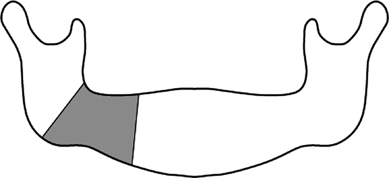
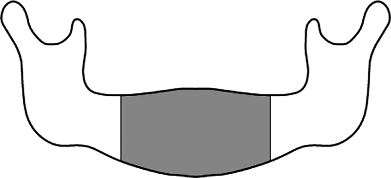
- Brown J S, Barry C, Ho M, Shaw R. A new classification for mandibular defects after oncological resection. Lancet Oncol. 2016;17(01):e23–e30. - PubMed
-
- Marx R E, Cillo J E, Jr, Broumand V, Ulloa J J. Outcome analysis of mandibular condylar replacements in tumor and trauma reconstruction: a prospective analysis of 131 cases with long-term follow-up. J Oral Maxillofac Surg. 2008;66(12):2515–2523. - PubMed
-
- Fernandes R P, Yetzer J G. Reconstruction of acquired oromandibular defects. Oral Maxillofac Surg Clin North Am. 2013;25(02):241–249. - PubMed
-
- Schrag C, Chang Y M, Tsai C Y, Wei F C. Complete rehabilitation of the mandible following segmental resection. J Surg Oncol. 2006;94(06):538–545. - PubMed
