. 2019 Feb 26:10:147.
doi: 10.3389/fphys.2019.00147. eCollection 2019.

MSP-RON Signaling Is Activated in the Transition From Pancreatic Intraepithelial Neoplasia (PanIN) to Pancreatic Ductal Adenocarcinoma (PDAC)

Affiliations

MSP-RON Signaling Is Activated in the Transition From Pancreatic Intraepithelial Neoplasia (PanIN) to Pancreatic Ductal Adenocarcinoma (PDAC)

Ce Li et al. Front Physiol. .

Abstract

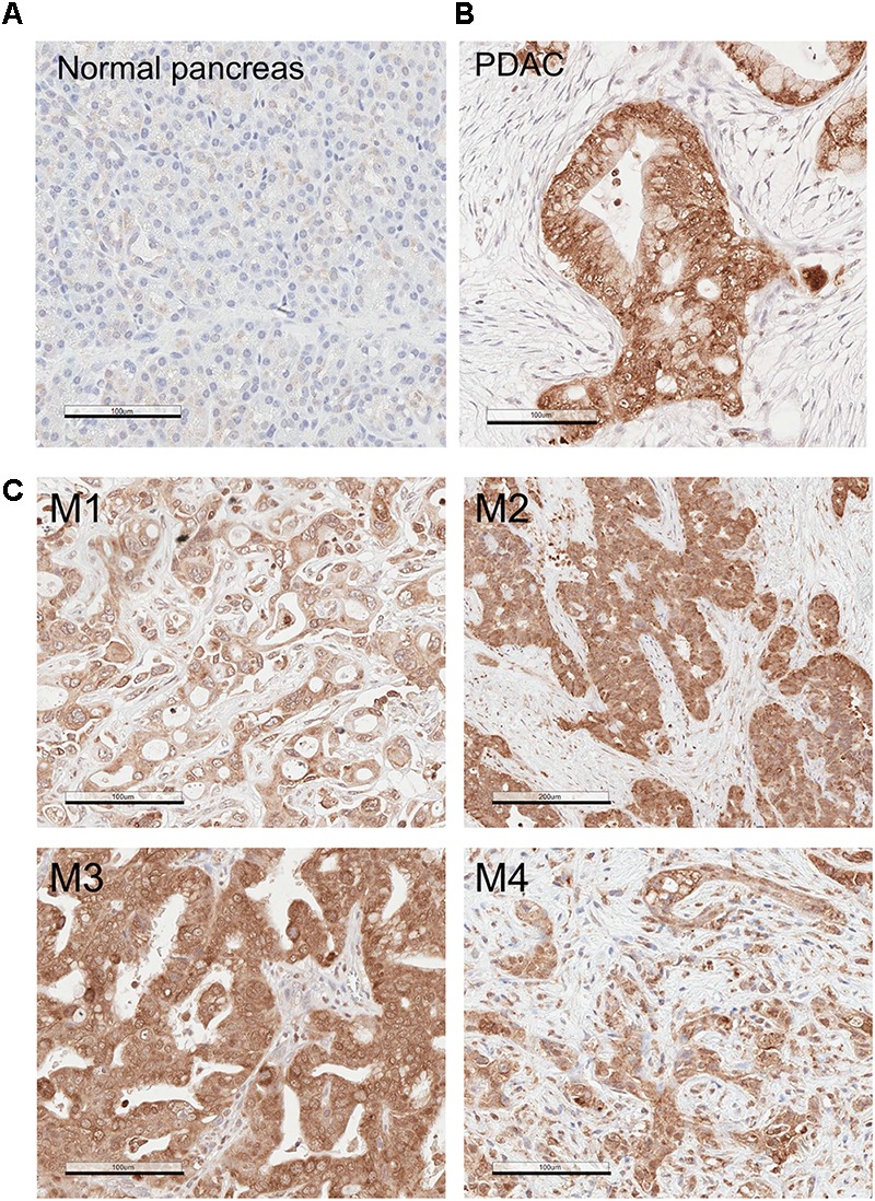
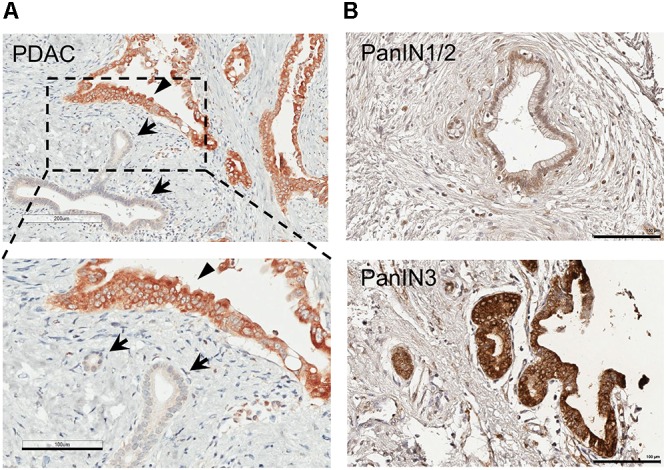
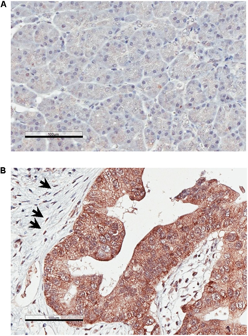
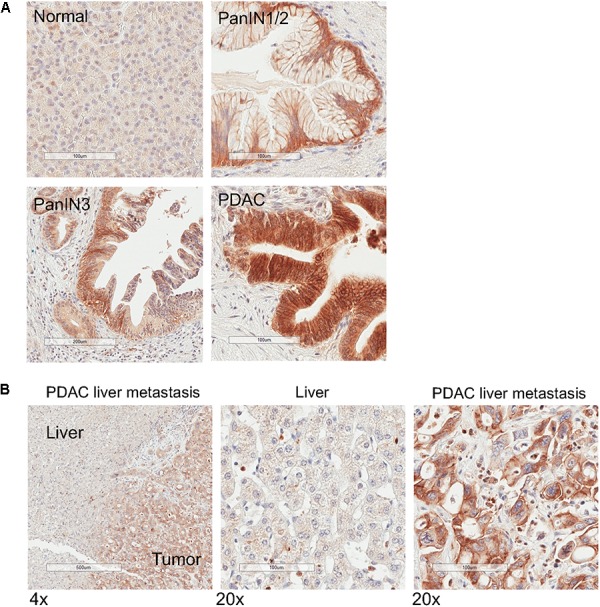
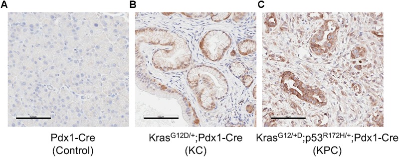
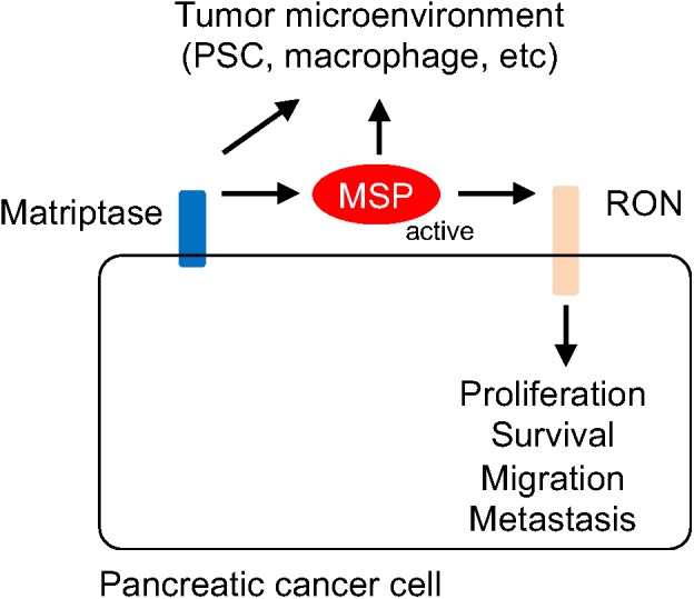
Pancreatic ductal adenocarcinoma (PDAC) is among the deadliest epithelial malignancies and remains difficult to treat. Pancreatic intraepithelial neoplasias (PanINs) represent the majority of the pre-cancer lesions in the pancreas. The PDAC microenvironment consists of activated pancreatic stellate cells (PSCs) and immune cells, which are thought to contribute to neoplastic transformation. However, the signaling events involved in driving the transition from the neoplastic precursor to the more advanced and aggressive forms in the pancreas are not well understood. Recepteur d'Origine Nantais (RON) is a c-MET family receptor tyrosine kinase that is implicated in playing a role in cell proliferation, migration and other aspects of tumorigenesis. Macrophage stimulating protein (MSP) is the ligand for RON and becomes activated upon proteolytic cleavage by matriptase (also known as ST14), a type II transmembrane serine protease. In the current study, by immunohistochemistry (IHC) analysis of human pancreatic tissues, we found that the expression levels MSP and matriptase are drastically increased during the transition from the preneoplastic PanIN stages to the more advanced and aggressive PDAC. Moreover, RON is highly expressed in both PDAC and in cancer-associated stellate cells. In contrast, MSP, RON, and matriptase are expressed at low levels, if any, in normal pancreas. Our study underscores an emerging role of MSP-RON autocrine and paracrine signaling events in driving malignant progression in the pancreas.

Keywords: MSP/MST1; RON/MST1R; matriptase; metastasis; pancreas; pancreatic ductal adenocarcinoma; pancreatic intraepithelial neoplasia; stellate cell.


Figures

FIGURE 1
FIGURE 1
MSP expression is upregulated in Pancreatic ductal adenocarcinoma (PDAC) primary tumors and liver metastasis. Immunohistochemistry (IHC) analysis of human tissues using anti-MSP antibody. (A) Normal pancreas; (B) PDAC; (C) Pancreatic cancer metastasis to the liver. Magnification: 20×; Scale bar: 100 μm.
FIGURE 2
FIGURE 2
MSP expression in human pancreatic cancer tissues and tissue micro array (TMA). IHC staining of human pancreatic tissues using anti-MSP antibody. (A) PDAC. Note that MSP can be detected in cancer cells (arrowheads) but not in normal or un transformed ductal epithelial cells (arrows). (B) Representative images of TMA with PanIN1/2 or 3. Magnification: 20×; Scale bar: 100 μm.
FIGURE 3
FIGURE 3
Recepteur d’Origine Nantais (RON) expression is increased in PDAC. IHC analysis of human pancreatic specimens using anti-RON antibody. (A) Normal pancreas; (B) PDAC. The arrows indicate RON-positive stellate cells. Magnification: 20×; Scale bar: 100 μm.
FIGURE 4
FIGURE 4
Matriptase expression is upregulated in PanIN, primary PDAC and liver metastasis. IHC analysis of normal human pancreatic tissues using anti-matriptase antibody. Representative staining images are shown. (A) Normal pancreas, PanIN1/2, PanIN 3, and PDAC; (B) Specimen of pancreatic cancer liver metastasis. The areas of the tumor and the adjacent tumor are indicated. Magnification: 4× (left panel) or 20×; Scale bar: 500 (left panel) or 100 μm.
FIGURE 5
FIGURE 5
MSP expression in mouse pancreatic tissues. IHC analysis of mouse pancreatic tissues using anti-MSP antibody. (A) control (Pdx1-Cre); (B) KC mice (LSL-KRASG12D/+; Pdx1-Cre); or (C) KPC mice (LSL-KRASG12D/+; LSL-p53R172H/+; Pdx1-Cre). Magnification: 20×; Scale bar: 100 μm.
FIGURE 6
FIGURE 6
Schematic representation of the mechanism by which the matriptase-MSP-RON signaling axis modulates pancreatic cancer progression. Matriptase mediates maturation and activation of MSP, which in turn stimulates RON-modulated signaling events. It is conceivable that Matriptase and MSP may also contribute to remodeling of the tumor microenvironment, through regulating the functions of the stellate cell, the macrophage, or other immune cells.

References

    1. Almoguera C., Shibata D., Forrester K., Martin J., Arnheim N., Perucho M. (1988). Most human carcinomas of the exocrine pancreas contain mutant c-K-ras genes. Cell 53 549–554. 10.1016/0092-8674(88)90571-5 - DOI - PubMed
    1. Apte M., Pirola R., Wilson J. (2009a). New insights into alcoholic pancreatitis and pancreatic cancer. J. Gastroenterol. Hepatol. 24(Suppl. 3), S51–S56. 10.1111/j.1440-1746.2009.06071.x - DOI - PubMed
    1. Apte M. V., Pirola R. C., Wilson J. S. (2009b). Pancreas: alcoholic pancreatitis–it’s the alcohol, stupid. Nat. Rev. Gastroenterol. Hepatol. 6 321–322. 10.1038/nrgastro.2009.84 - DOI - PubMed
    1. Apte M. V., Haber P. S., Darby S. J., Rodgers S. C., Mccaughan G. W., Korsten M. A., et al. (1999). Pancreatic stellate cells are activated by proinflammatory cytokines: implications for pancreatic fibrogenesis. Gut 44 534–541. 10.1136/gut.44.4.534 - DOI - PMC - PubMed
    1. Ardito C. M., Gruner B. M., Takeuchi K. K., Lubeseder-Martellato C., Teichmann N., Mazur P. K., et al. (2012). EGF receptor is required for KRAS-induced pancreatic tumorigenesis. Cancer Cell 22 304–317. 10.1016/j.ccr.2012.07.024 - DOI - PMC - PubMed