Review
. 2019 Feb 26:10:286.
doi: 10.3389/fmicb.2019.00286. eCollection 2019.

Host Lipids in Positive-Strand RNA Virus Genome Replication

Affiliations
Review

Host Lipids in Positive-Strand RNA Virus Genome Replication

Zhenlu Zhang et al. Front Microbiol. .

Abstract

Membrane association is a hallmark of the genome replication of positive-strand RNA viruses [(+)RNA viruses]. All well-studied (+)RNA viruses remodel host membranes and lipid metabolism through orchestrated virus-host interactions to create a suitable microenvironment to survive and thrive in host cells. Recent research has shown that host lipids, as major components of cellular membranes, play key roles in the replication of multiple (+)RNA viruses. This review focuses on how (+)RNA viruses manipulate host lipid synthesis and metabolism to facilitate their genomic RNA replication, and how interference with the cellular lipid metabolism affects viral replication.

Keywords: lipid metabolism; membrane association; phospholipids; positive-strand RNA virus; viral RNA replication.


Figures
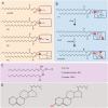
Figure 1
Figure 1
Structures of major lipids. (A,B) Phospholipid structure representations. Boxed areas indicate the different headgroups among various phospholipids. (C) Structure of sphingolipids. X represents different headgroups of different sphingolipids. (D) Basic structure of sterol (Left) and the structure of cholesterol (Right). PA, phosphatidic acid; PE, phosphatidylethanolamine; PC, phosphatidylcholine; PI, phosphatidylinositol; PI4P, phosphatidylinositol-4-phosphate; PI(4,5)P2, phosphatidylinositol-4,5-bisphosphate; PI4K, Phosphatidylinositol-4 kinase; PI4P5K, phosphatidylinositol 4-phosphate 5-kinase; Cer, ceramide; SM, sphingomyelin; GSL, glycosphingolipid.
Figure 2
Figure 2
Different (+)RNA viruses explore specific cellular organelle membranes for VRCs assembly and genome replication. (A) Both ER and Golgi membranes are used for the formation of Poliovirus VRCs. The 3D model indicates the single-membrane tubules formed at early stage of poliovirus infection. (B) HCV utilizes ER membranes to form double-membrane vesicles (DMVs) as VRCs. (C) DENV replicates in association with ER membranes. ER membranes are in yellow, VRCs are in light brown. Note virions (in red) are produced near VRCs. (D) BMV invaginates the outer perinuclear ER membrane to assemble VRCs. Nuc: nucleus; cyto: cytoplasm. (E) TBSV replicates in association with the peroxisomal membranes and VRCs (blue) are formed inside multivesicular bodies (MVB, yellow membranes). (F) FHV invaginates the outer mitochondiral membranes (blue) to build VRCs (White). Arrows point to VRCs. (A–F) are reproduced with permission from Belov et al. (2012) in (A); Romero-Brey et al. (2012) in (B); Welsch et al. (2009) in (C); Schwartz et al. (2002) in (D); Fernandez de Castro et al. (2017) in (E); and Kopek et al. (2007) in (F).
Figure 3
Figure 3
The distribution of major lipids in host organelle membranes. Different colors of circles represent different lipid classes, and the size of circles represent the percentage of one class of lipid to the total phospholipids, as modified from van Meer et al. (2008). CL, cardiolipin; PA, phosphatidic acid; PC, phosphatidylcholine; PE, phosphatidylethanolamine; PS, phosphatidylserine; PI, phosphatidylinositol; SM, sphingomyelin; St, sterol; StE, steryl ester; TAG, triacylglycerol.
Figure 4
Figure 4
Models for the formation of membrane curvature and viral replication complexes. (A) The insertion of certain lipids with cone or inverted-cone shape generates negative or positive membrane curvature, respectively. (B,C) Models for the formation of invagination- and protrusion-type replication complexes. In the protrusion-type model, the VRC on the right represents the double-membrane vesicle (DMV). PC, phosphatidylcholine; PI, phosphatidylinositol; PI4P, phosphatidylinositol-4-phosphate; PI(4,5)P2, phosphatidylinositol-4,5-bisphosphate; PI(3,4,5)P3, phosphatidylinositol-3,4,5-trisphosphate; PA, phosphatidic acid; SM, sphingomyelin; PE, phosphatidylethanolamine; UFA, unsaturated fatty acid; Cer, ceramide; VRC, virus replication complex.
Figure 5
Figure 5
Pathways for the biosynthesis of major lipids. Key enzymes in pathways or those that are recruited by viruses are listed. The conversion from PE to PC is catalyzed by Cho2p and Opi3p in yeast but PLMT in mammals. Yeast or plant Pah1p or mammalian lipin converts DAG to PA. Gro-3-P, glycerol-3-phosphate; Lyso-PA, lysophosphatidic acid; PA, phosphatidic acid; CDP-DAG, cytidine diphosphate diacylglycerol; PI, phosphatidylinositol; PI4K, Phosphatidylinositol-4 kinase; PI4P, phosphatidylinositol-4-phosphate; PI4P5K, phosphatidylinositol 4-phosphate 5-kinase; PI(4,5)P2, phosphatidylinositol-4,5-bisphosphate; PS, phosphatidylserine; PE, phosphatidylethanolamine; PC, phosphatidylcholine; Etn, ethanolamine; Cho, choline; DAG, diacylglycerol; TAG, triacylglycerol; StE, steryl ester; ErgE, ergosterol ester; Acyl-CoA, acyl-coenzyme A; FFA, free fatty acid; FA, fatty acid; FASN, fatty acid synthase; HMG-CoA, 3-hydroxy-3-methylglutaryl-coenzyme A; SPT, serine palmitoyltransferase; 3-KDS, 3-ketodihydrosphingosine; GlcCer, glucosylceramide; LacCer, lactosylceramide; SM, sphingomyelin; SL, sphingolipid; GSL, glycosphingolipid; FAPP2, four-phosphate adaptor protein 2.

Similar articles

Cited by

References

    1. Adachi-Yamada T., Gotoh T., Sugimura I., Tateno M., Nishida Y., Onuki T., et al. . (1999). De novo synthesis of sphingolipids is required for cell survival by down-regulating c-Jun N-terminal kinase in Drosophila imaginal discs. Mol. Cell. Biol. 19, 7276–7286. 10.1128/MCB.19.10.7276 - DOI - PMC - PubMed
    1. Ahlquist P. (2006). Parallels among positive-strand RNA viruses, reverse-transcribing viruses and double-stranded RNA viruses. Nat. Rev. Microbiol. 4, 371–382. 10.1038/nrmicro1389 - DOI - PMC - PubMed
    1. Aizaki H., Lee K. J., Sung V. M. H., Ishiko H., Lai M. M. C. (2004). Characterization of the hepatitis C virus RNA replication complex associated with lipid rafts. Virology 324, 450–461. 10.1016/j.virol.2004.03.034 - DOI - PubMed
    1. Aktepe T. E., Pham H., Mackenzie J. M. (2015). Differential utilisation of ceramide during replication of the flaviviruses West Nile and dengue virus. Virology 484, 241–250. 10.1016/j.virol.2015.06.015 - DOI - PubMed
    1. Albert B., Johnson A., Lewis J., Raff M., Roberts K., Walter P. (2002). The Lipid Bilayer in Molecular Biology of the Cell. New York, NY: Garland Science.

LinkOut - more resources